
Image
|
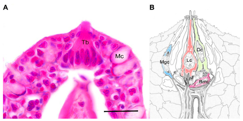
Figure Caption
Fig. 1
Taste bud. (A) Histological section of a single taste bud protruding from the pluristratified epithelium of the oropharyngeal cavity. Hematoxilin-eosin staining; (B) Schematic representation of the ultrastructural organization of fish taste buds (modified from Hansen et al. 2002). Abbreviations: Tb = taste bud; Mc = mucous cell; Dc = dark cell (green); Lc = light cell (red); Bmc = basal cell (pink); Mgc = marginal cell (blue). Scale bar = 20 µm.
Acknowledgments
This image is the copyrighted work of the attributed author or publisher, and
ZFIN has permission only to display this image to its users.
Additional permissions should be obtained from the applicable author or publisher of the image.
Full text @ Animals (Basel)