
IMAGE
Fig. 4
- ID
- ZDB-IMAGE-220701-23
- Publication
- Ren et al., 2022 - Cryo-EM structure of the heptameric calcium homeostasis modulator 1 channel
- All Figures
- Figures for Ren et al., 2022
Image
|
Figure Caption
Fig. 4
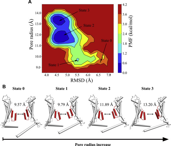
Figure 4. The updown motion of the N-helix increases the pore diameter. A, two-dimensional PMF profile of the RMSD of the P1 N-helix versus the pore radius in the heptameric channel. The initial structure and three low-energy conformational states during supervised MD simulations are labeled state 0, state 1, state 2, and state 3. B, representative structures of P4 and P7 in state 0, state 1, state 2 and state 3 of the heptameric channel. Two opposing protomers of the heptamer are shown in cartoon. PMF, potential of mean force.
Acknowledgments
This image is the copyrighted work of the attributed author or publisher, and
ZFIN has permission only to display this image to its users.
Additional permissions should be obtained from the applicable author or publisher of the image.
Full text @ J. Biol. Chem.