
IMAGE
Fig. 1
- ID
- ZDB-IMAGE-220701-20
- Publication
- Ren et al., 2022 - Cryo-EM structure of the heptameric calcium homeostasis modulator 1 channel
- All Figures
- Figures for Ren et al., 2022
Image
|
Figure Caption
Fig. 1
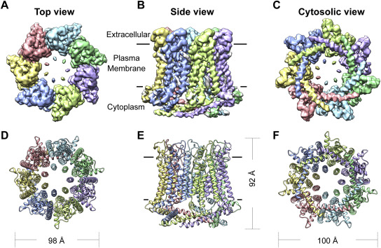
Figure 1. The overall architecture of heptamer+LCH. The surface (A) and cartoon (D) representations of the cryo-EM density map of heptameric drCALHM1 viewed from the extracellular side of the membrane. The seven subunits are represented by different colors. The surface (B) and cartoon (E) representations of the cryo-EM density map of heptamer+LCH viewed parallel to the membrane. The lipid-like density is gray. The surface (C) and cartoon (F) representations of the cryo-EM density map of heptamer+LCH viewed from the cytosolic side of the membrane. LCH, long C-terminal helix.
Acknowledgments
This image is the copyrighted work of the attributed author or publisher, and
ZFIN has permission only to display this image to its users.
Additional permissions should be obtained from the applicable author or publisher of the image.
Full text @ J. Biol. Chem.