
Fig. 1
- ID
- ZDB-IMAGE-220629-83
- Antibodies
- Publication
- Dash et al., 2022 - Nucleolin loss of function leads to aberrant Fibroblast Growth Factor signaling and craniofacial anomalies
- All Figures
- Figures for Dash et al., 2022
|
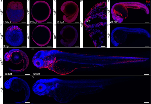
Fig. 1
Ncl expression during zebrafish development. (A) During embryogenesis, Nucleolin (Ncl, red) was ubiquitously expressed in the cytoplasm of four-cell stage wild-type embryo at 1.5 hpf as observed by immunostaining. (B) Similarly, 3 hpf embryos also had ubiquitous cytoplasmic expression of Nucleolin. (C,C′) At 12 hpf, ncl+/+ and ncl−/− embryos exhibited similar Nucleolin expression in the nucleus and cytoplasm in most cells of the embryos. (D-D″) At 18 hpf, the expression of Nucleolin in ncl+/+ embryos was ubiquitous and was confined to the nucleus (D″). In the ncl−/− embryos, the expression pattern of Nucleolin was similar to that of wild-type embryos; however, the expression levels were significantly lower than that of the wild type. (E,E′) By 24 hpf, the expression of Nucleolin was still ubiquitous, with higher levels in the eye and the midbrain-hindbrain boundary in ncl+/+ embryos, whereas it was absent in ncl−/− embryos. (F) At 36 hpf, the expression of Nucleolin became specific to the craniofacial region in the pharyngeal arches as well as the eye. (G) In 72 hpf (3 dpf) wild-type zebrafish, Nucleolin was highly expressed in the jaw of the embryo. (F′,G′) In the ncl−/− mutants, there was no expression of Nucleolin. n=15 for each panel. The experiment was performed three times. NT, neural tube. Scale bars: 35 µm (A,B); 70 µm (C,C′); 140 µm (D,D′); 50 µm (D″); 250 µm (E,E′); 300 µm (F,F′,G,G′).