
|
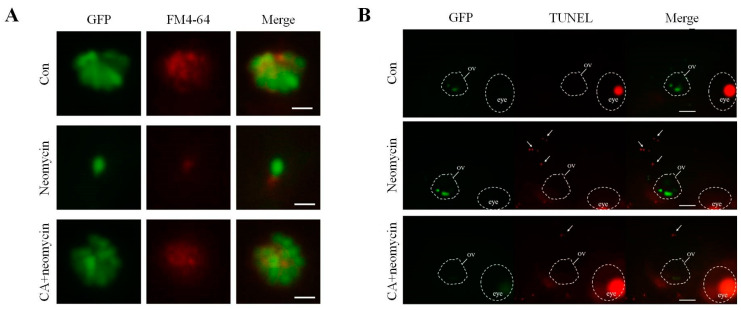
Fig. 3
Evaluation of CA protected against neomycin-induced apoptosis and mechanotransduction channel impairment. (A) Fluorescence photograph of hair cells in neuromasts of 7-dpf transgenic zebrafish larvae treated with 12.5 μM of neomycin for 0.5 h only or pre-treated with 5 μM CA for 2 h. Labeling FM4-64 fluorescence dye that passes through hair’s h mechanotransduction channel was market d as a red-color signal. A comparison of the signal intensity between untreated control, neomycin, and neomycin pre-treated with CA for 2 h showed that CA prevented neomycin-induced mechanotransduction channel impairment. Scale bar: 10 μm. (B) Apoptotic hair cells were marked as light-respotsot (middle le) in the anterior region of the lateral line system after TUNEL staining. The location of the oval window (ov) and eye a were marked by the white circle. Comparison of TUNEL-positive signal intensity between untreated control, neomycin, and neomycin pre-treated with CA for 2 h showed that CA prevented neomycin-induced apoptosis of TUNEL-positive cells. Scale bar: 100 μm.