
|
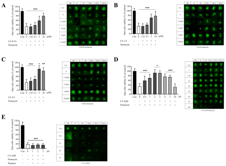
Fig. 2
CA protected against neomycin-induced lateral line hair cell loss. (A) Transgenic zebrafish larvae were fixed and photographed using a fluorescence microscope. 7-dpf transgenic zebrafish larvae were treated with 12.5 μM of neomycin only for 0.5 h or pre-treated with CA (0, 1.25, 2.5, 5, and 10 μM) for 0.5 h, (B) 1 h, and (C) 2 h, respectively. (D) 7-dpf transgenic zebrafish larvae were treated with 12.5 μM of neomycin only for 0.5 h or pretreated with 5 μM CA for different times (0, 0.5, 1, 2, 3, 4, 5, and 24 h). (E) 7-dpf transgenic zebrafish larvae were pretreated with 5 μM of CA for 1, 2, and 3 h followed by CA washout, and subsequently 0.5 h of 12.5 μM neomycin treatment. Fluorescence micrographs of lateral line hair cells from neuromasts (O, OC, MI, PLL1, PLL3, and PLL4) (right panel) were analyzed and the quantitative result of viable hair cells was present as a percentage of the untreated control (left panel). All values of the experimental groups were presented as mean ± SD. ** p < 0.01, *** p < 0.001, and ns, no significant difference as compared with untreated control.