
|
Fig. 1
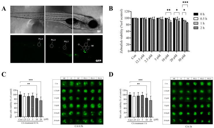
Effects of CA on transgenic zebrafish larvae and hair cell viability. (A) Fluorescence micrograph of a 7-dpf transgenic zebrafish larvae. The analyzed lateral line hair cells in zebrafish are marked by a white circle: otic (O), occipital (OC), middle (MI), and posterior lateral line (PLL1, PLL3, and PLL4). Scale bar: 100 μm. (B) 7-dpf transgenic zebrafish larvae were treated with CA (0, 1.25, 2.5, 5, 10, 20, and 30 μM) for different time (0.5, 1, and 2 h). After the exposure period, viable fish were counted and presented as a percentage of the untreated control. (C) Lateral line hair cells were treated with CA (0, 1.25, 2.5, 5, 10, and 20 μM) for 0.5 h and (D) 1 h. Fluorescence micrographs of lateral line hair cells from neuromasts (O, OC, MI, PLL1, PLL3, and PLL4) (right panel) were analyzed and the quantitative result of viable hair cells was present as a percentage of the untreated control (left panel). All values of the experimental groups were presented as mean ± SD. * p < 0.05, ** p < 0.01, and *** p < 0.001 as compared with untreated control. CA, CA; O, otic; OC, occipital; MI, middle; and PLL, posterior lateral line.