
Fig. 6 - supplement 1
- ID
- ZDB-IMAGE-220617-10
- Publication
- Lai et al., 2022 - DNA-damage induced cell death in yap1;wwtr1 mutant epidermal basal cells
- All Figures
- Figures for Lai et al., 2022
|
Fig. 6 - supplement 1
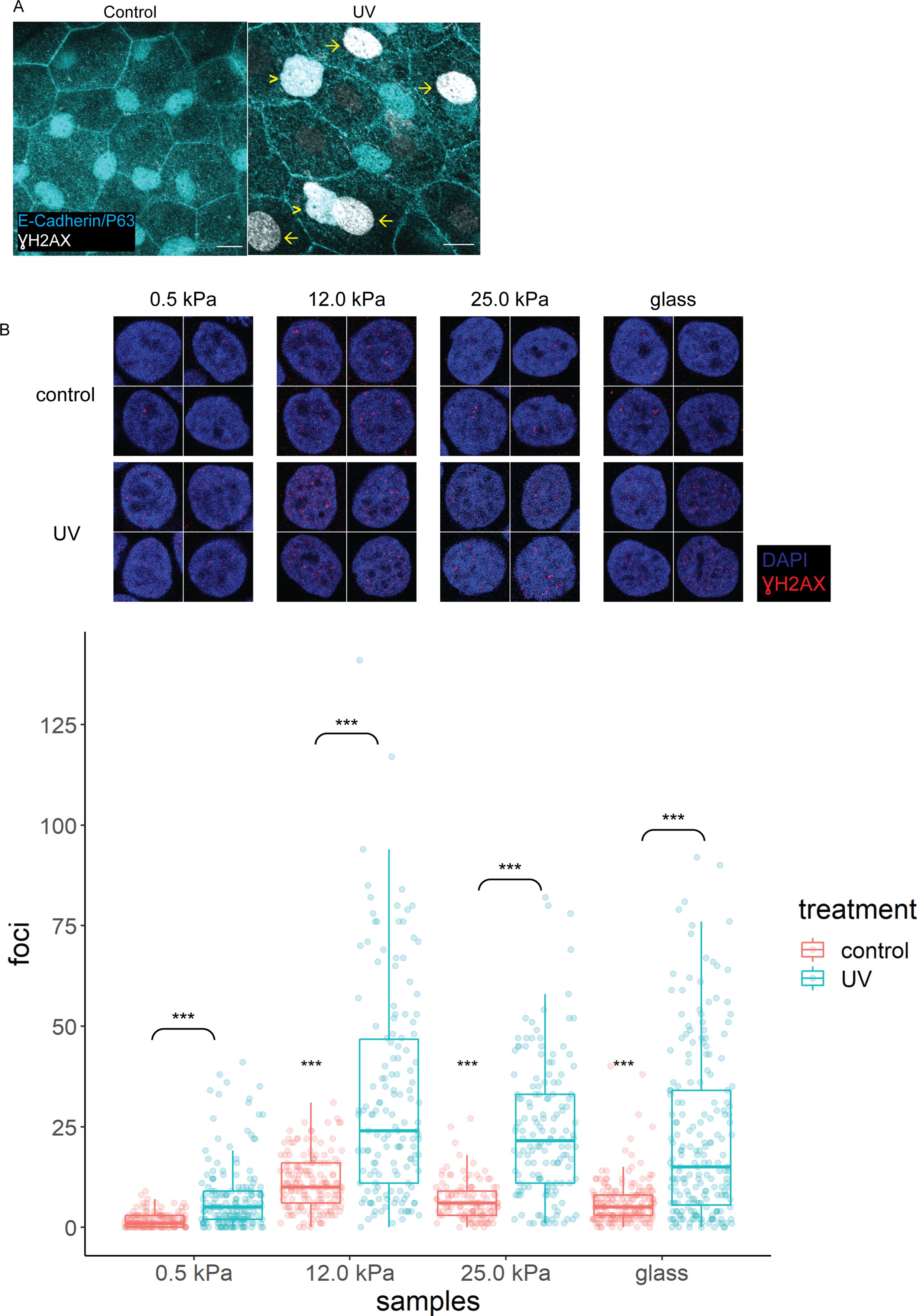
(A) Control (n=5) and UV-treated (n=4) head epidermal cells of zebrafish embryos stained with γH2AX and epidermal markers (E-cadherin and P63). P63 is a marker for epidermal basal cells (EBCs). EBCs (yellow arrowheads) and peridermal cells (yellow arrows) exposed to UV exhibit pan-nuclear γH2AX. (B) Selected nuclei of control and UV-treated HaCaT cells cultured on 0.5, 12, 25 kPa hydrogels and glass, and boxplots of the number of γH2AX foci in these nuclei. t-Tests on control groups were compared against control HaCaT cells cultured on 0.5 kPa hydrogel. t-Tests on UV groups were compared against their respective controls (brackets). Data were collected from three independent experiments. ***p<0.001 adjusted for multiple testing.
γH2AX in zebrafish head epidermis and HaCaT cells exposed to UV.