
Image
|
Figure Caption
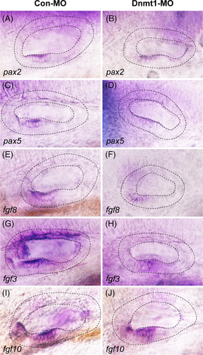
Fig. 8
Changes in the expression of otic placode marker genes after Dnmt1 knockdown. In situ staining of pax2 and pax5 are all downregulated in Dnmt1-MO embryos compared to the controls by WISH at 48 hpf. The black dotted lines outline the otic vesicle. The Fgf signalling ligands fgf8, fgf3 and fgf10 are in lower expression in Dnmt1-MO embryos compared to the controls at 48 hpf
Acknowledgments
This image is the copyrighted work of the attributed author or publisher, and
ZFIN has permission only to display this image to its users.
Additional permissions should be obtained from the applicable author or publisher of the image.
Full text @ Cell Prolif.