
IMAGE
Fig. 5
- ID
- ZDB-IMAGE-220607-32
- Publication
- Varela et al., 2022 - Cdkl5 mutant zebrafish shows skeletal and neuronal alterations mimicking human CDKL5 deficiency disorder
- All Figures
- Figures for Varela et al., 2022
Image
|
Figure Caption
Fig. 5
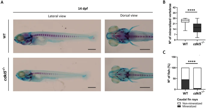
Skeletal development of cdkl5sa21938 mutants with 14 dpf. (A) Representative images of the wild-type (WT) and homozygous (cdkl5-/-) larvae double-stained with alcian blue and alizarin red. Scale bar = 0.5 mm. (B) Number of mineralized vertebrae in cdkl5−/− (n = 58) and WT (n = 54) larvae. Values are represented as median with interquartile range. Statistical analysis was performed using Mann–Whitney. (C) Percentage of fish with mineralized caudal fin rays. Statistical analysis was performed using the Chi-square test. **** indicate p < 0.0001.
Figure Data
Acknowledgments
This image is the copyrighted work of the attributed author or publisher, and
ZFIN has permission only to display this image to its users.
Additional permissions should be obtained from the applicable author or publisher of the image.
Full text @ Sci. Rep.