
IMAGE
Fig. 1
- ID
- ZDB-IMAGE-220502-10
- Genes
- Publication
- Seese et al., 2021 - Genetic disruption of zebrafish mab21l1 reveals a conserved role in eye development and affected pathways
- All Figures
- Figures for Seese et al., 2021
Image
|
Figure Caption
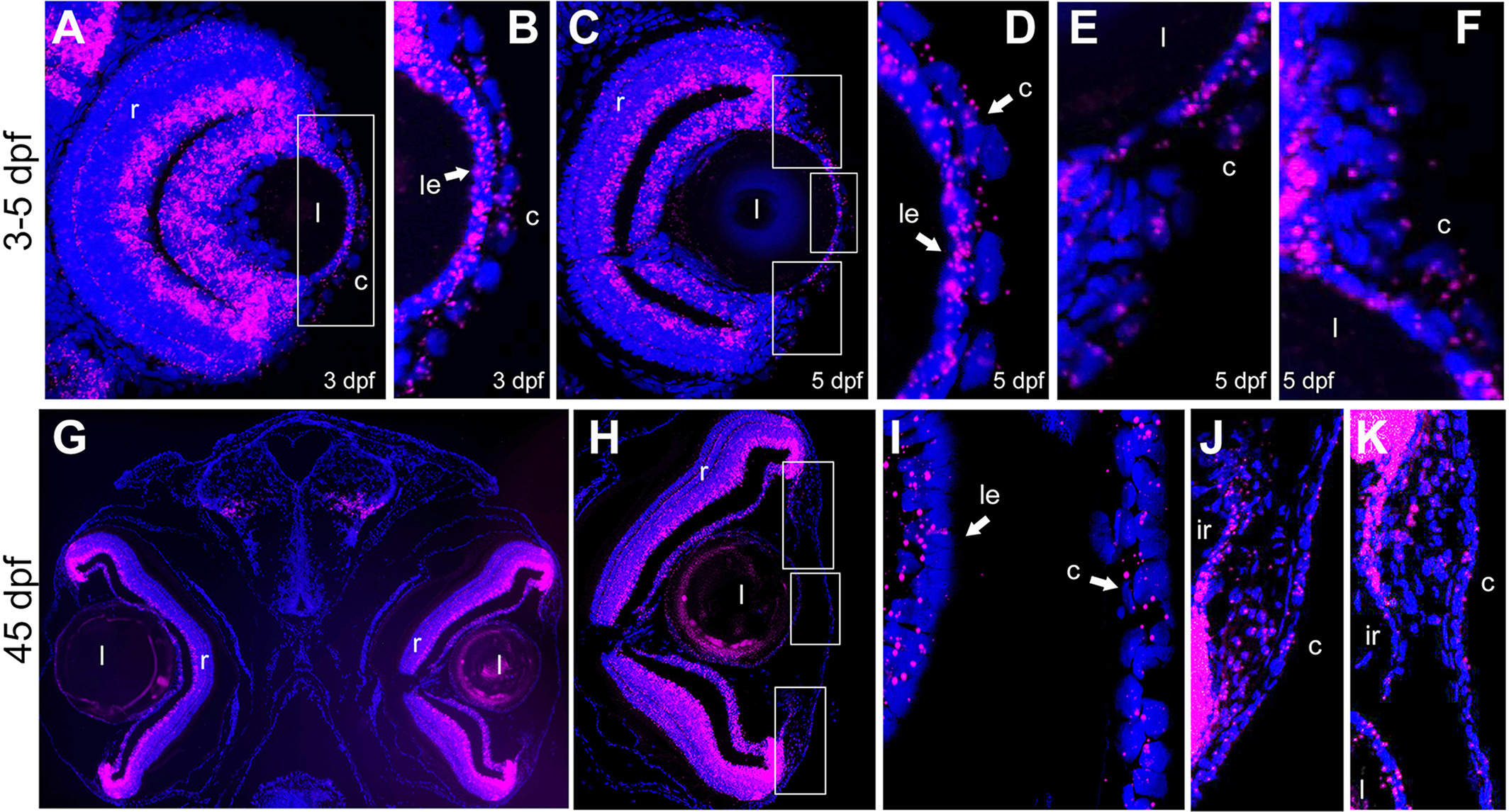
Fig. 1 Expression pattern of mab21l1 in 3-, 5-, and 45-dpf zebrafish eye. RNA-scope analysis of mab21l1 (magenta) in 3-dpf, A and B, 5-dpf, C-F, and 45-dpf, G-K, transverse sections. At all stages, mab21l1 is expressed in the cornea (c) (both central, B, D, I, and peripheral, E, F, J, K), lens (l) (specifically, anterior lens epithelium [le]), and retina (r), A-K, as well as dorsal and ventral iris (ir) and iridocorneal angle at 45-dpf, J, K. DAPI staining indicates cell nuclei (blue). The boxed regions in, A, C, and H are shown in enlarged forms in, B, D-F, and I-K, respectively
Figure Data
Acknowledgments
This image is the copyrighted work of the attributed author or publisher, and
ZFIN has permission only to display this image to its users.
Additional permissions should be obtained from the applicable author or publisher of the image.
Full text @ Dev. Dyn.