Image
Figure Caption
Figure 1
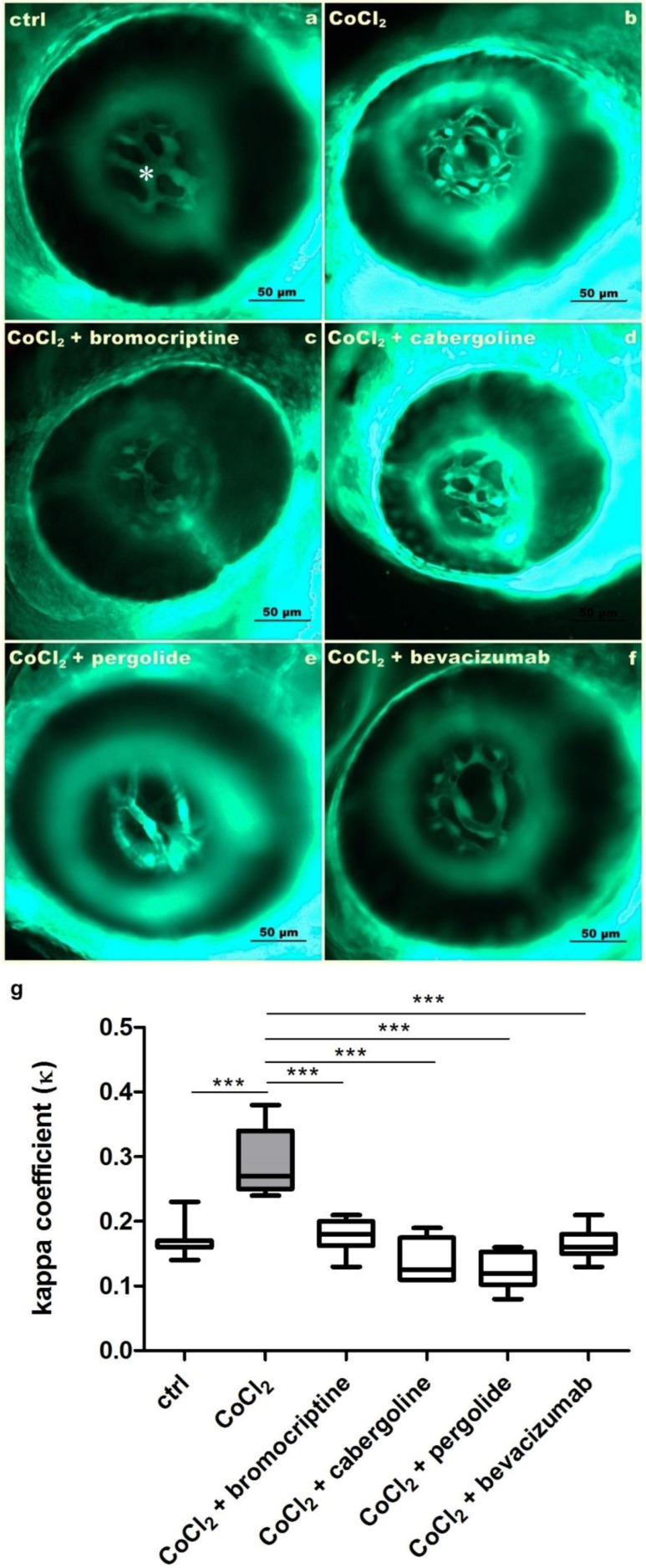
Figure 1. A set of microphotographs and a graph documenting inhibition of 5 mM CoCl2-derived increased angiogenesis of hyaloid-retinal vessels (HRVs) (asterisk) resulting from co-treatment with dopaminergic agonists in 3 dpf Tg(Fli-1:EGFP) zebrafish larvae. (a) The control untreated larva; HRVs are marked with *. (b) 5 mM CoCl2 exposure resulted in increased HRVs branching. (c–g) Co-treatment with 2.5 µM/L bromocriptine, 2.5 µM/L cabergoline, 2.5 µM/L pergolide and 2.5 µM/L bevacizumab, respectively, resulted in significant inhibition of HRVs abnormal branching. (g) The graph presenting the kappa coefficient (ĸ) values (ratio of HRVs area to eyeball area) in investigated groups. (one-way ANOVA, GraphPad Prism 5, *** p < 0.001).
Acknowledgments
This image is the copyrighted work of the attributed author or publisher, and
ZFIN has permission only to display this image to its users.
Additional permissions should be obtained from the applicable author or publisher of the image.
Full text @ Cells

