
|
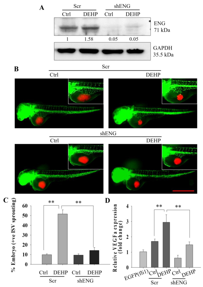
Figure 3 Representative and quantitative results showing that DEHP-induced endoglin-controlled VEGFA-mediated angiogenesis in vivo. (A) Prolonged DEHP treatment upregulated endoglin expression in MDA-MB-231 cells, and shENG treatment depleted endoglin expression in control and DEHP-exposed MDA-MB-231 cells. (B) Endoglin knockdown reduced DEHP-induced SIV sprouting in Tg (fli1: EGFP) zebrafish embryos at 24 hpi. Scale bar =1000 µm. (C) Quantitative results of angiogenesis showing a reduced number of zebrafish embryos showing SIV sprouting following endoglin knockdown (n = 50). (D) Depletion of VEGFA mRNA levels in zebrafish embryos following endoglin knockdown cell xenografts of DEHP-exposed MDA-MB-231 cells; ** p < 0.001.