
IMAGE
Fig 7
- ID
- ZDB-IMAGE-220331-42
- Genes
- Publication
- Reuter et al., 2022 - Identification of an evolutionarily conserved domain in Neurod1 favouring enteroendocrine versus goblet cell fate
- All Figures
- Figures for Reuter et al., 2022
Image
|
Figure Caption
Fig 7
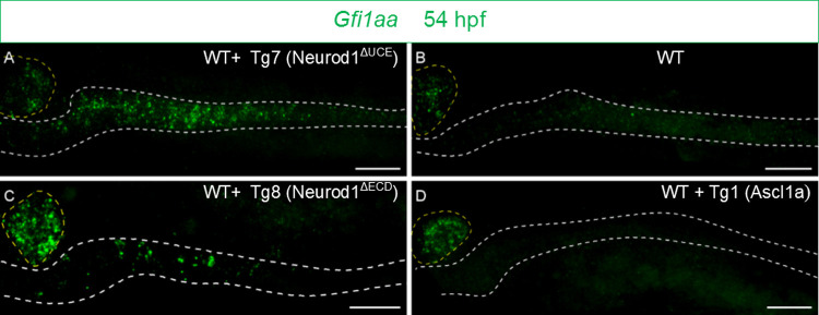
FISH performed with the
Figure Data
Acknowledgments
This image is the copyrighted work of the attributed author or publisher, and
ZFIN has permission only to display this image to its users.
Additional permissions should be obtained from the applicable author or publisher of the image.
Full text @ PLoS Genet.