
Image
|
Figure Caption
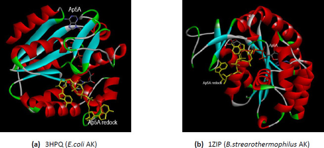
Figure 3.
The comparative interactions of the bacterial AKs with the analog substrate Ap5A after redocking. The redocked conformations are shown in yellow. (a) E.coli AK (PDB ID 3HPQ); (b) B.stearothermophilus AK (PDB ID 1ZIP).
Acknowledgments
This image is the copyrighted work of the attributed author or publisher, and
ZFIN has permission only to display this image to its users.
Additional permissions should be obtained from the applicable author or publisher of the image.
Full text @ ADMET DMPK