
|
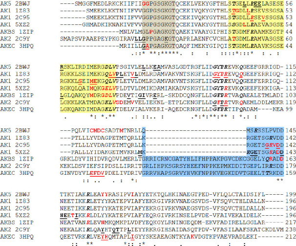
Figure 12.
Multiple sequence alignment of the AKs sequences. In gray background are marked the residues belong to Walker A motif – phosphate-binding loop or P-loop; in yellow background is marked the NMP binding region and the residues that belong to AMP binding region are italic-bolded; in blue background is marked the LID region; with red are marked the residues (van der Waals, conventional hydrogen bond or alkyl interactions) which interact with amantadine ionized form (with –NH3+ group); the residues which interact with amantadine unionized form (with –NH2 group) are bolded-underlined; the residues which interact with amantadine unionized and ionized are red and bolded-underlined; AKEC is AK from E.coli; AKBS is AK from B.stearothermophilus; with dot “.” are marked the semi-conservative replacements; with colon “:” are marked the conservative replacements; with “*” are marked the identities of the residues.