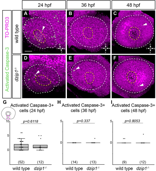
IMAGE

Fig 3

ID
ZDB-IMAGE-220318-4
Antibodies
Source
Figures for Nandamuri et al., 2022
Image
Figure Caption

Fig 3 Apoptotic cell death in the optic fissure is not aberrantly upregulated in the <italic toggle='yes'>dzip1</italic> mutant.

(A-F) Whole mount immunofluorescence in wild type (A-C) and dzip1ts294e mutants (D-F) for cell death (green; activated Caspase-3) and nuclei (magenta; TO-PRO-3) at 24 hpf (A, D), 36 hpf (B, E), and 48 hpf (C, F). All images are lateral views of 3-dimensional renderings. White dashed lines mark the boundary of the eye; yellow dashed lines mark the lens. Examples of cells labeled with activated caspase-3 elsewhere within the eye are indicated (white arrowheads). (G-I) Quantification of total number of activated caspase-3-positive cells within the optic fissure in the eye at 24 hpf, 36 hpf, and 48 hpf. n (embryos) for each genotype shown at the base of the graph. P-values were calculated using Welch’s t-test (M-O). Scale bar: 50 μm. D, dorsal; V, ventral; N, nasal; T, temporal.

Figure Data
Acknowledgments
This image is the copyrighted work of the attributed author or publisher, and ZFIN has permission only to display this image to its users. Additional permissions should be obtained from the applicable author or publisher of the image. Full text @ PLoS One