
Image
|
Figure Caption
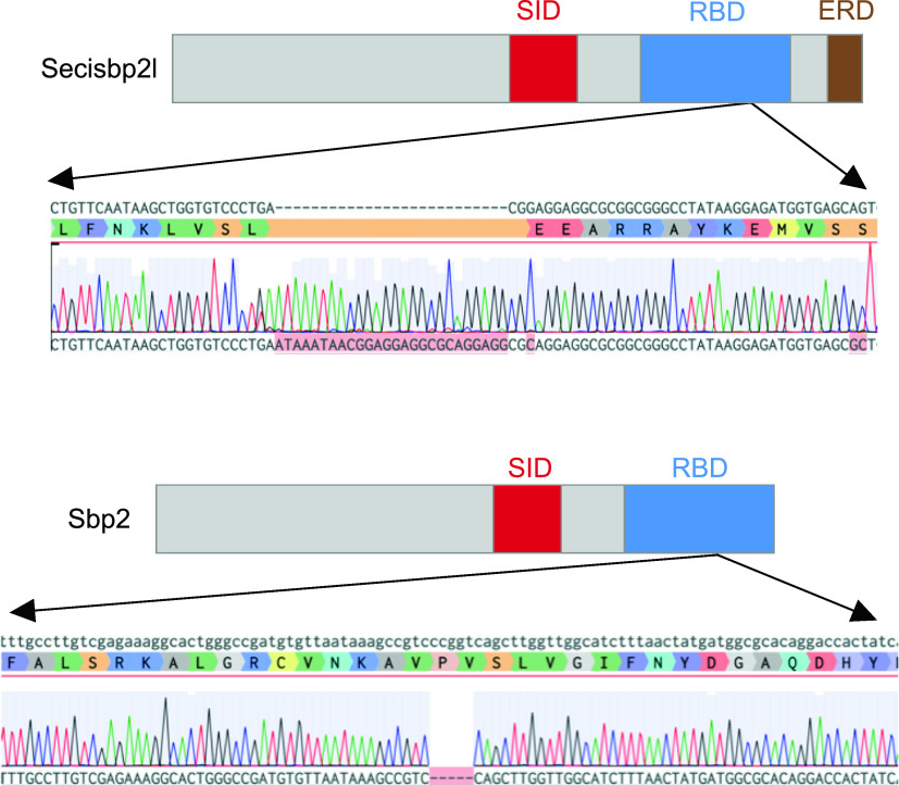
Figure 3.
CRISPR/Cas9 mutagenesis of the secisbp2l and sbp2 loci.
Domain diagrams illustrating the relative positions of the Sec incorporation domain (SID), RNA-binding domain (RBD) and the Glu-rich domain (ERD), and sequence analysis of secisbp2l and sbp2 mutations generated by CRISPR/Cas9 illustrating the 26-bp insertion for secisbp2l and the 5 bp deletion in sbp2.
Acknowledgments
This image is the copyrighted work of the attributed author or publisher, and
ZFIN has permission only to display this image to its users.
Additional permissions should be obtained from the applicable author or publisher of the image.
Full text @ Life Sci Alliance