IMAGE

Figure 3

ID
ZDB-IMAGE-220316-40
Source
Figures for Eaton et al., 2021
Image
Figure Caption

Figure 3

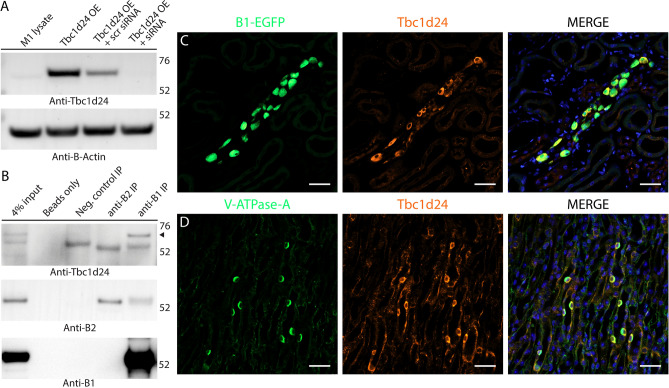
Tbc1d24 is expressed in mouse kidney intercalated cells (ICs) and co-immunoprecipitates with the kidney-specific B1 subunit of V-ATPase. (A) Validation of the commercial anti-Tbc1d24 antibodies, used in this study. Anti-Tbc1d24 western blot demonstrates that a band of the expected 63 kDa molecular mass is present in the lysate of M-1 cells overexpressing mouse Tbc1d24 (Tbc1d24 OE), but not in the lysate of M-1 cells co-transfected with both Tbc1d24-expressing plasmids and Tbc1d24-specific siRNA (Tbc1d24 OE + siRNA), confirming specificity of anti-Tbc1d24 antibodies. Anti-β-actin blot was used as a loading control. (B) Tbc1d24 co-immunoprecipitates with the kidney-enriched B1 subunit of the V-ATPase, but not with the ubiquitously expressed B2 subunit of V-ATPase in kidney. Proteins were co-immunoprecipitated using anti-B1 and anti-B2 antibodies from mouse kidney lysates and then analyzed by western blot, using anti-Tbc1d24 antibodies. The specific band of the expected 63 kDa molecular mass (arrowhead) is present only in the anti-B1 immunoprecipitation (anti-B1 IP) lane. Note, that the 50 kDa heavy chains of antibodies, used for immunoprecipitation, are detected in anti-B1 IP, anti-B2 IP and isotype control antibodies IP (neg. control IP) lanes, as expected. Anti-B1 and anti-B2 western blots are shown to confirm the successful immunoprecipitation of B1 and B2 subunits of V-ATPase. Anti-B1 antibodies co-immunoprecipitate the B2 subunit of V-ATPase, while anti-B2 antibodies do not co-immunoprecipitate the B1 subunit of V-ATPase, likely because there are many more homo B2/B2 complexes in the whole kidney, than hybrid B1/B2 complexes. This experiment was repeated three times with similar results. (C,D). Tbc1d24 is co-expressed with V-ATPase in mouse kidney intercalated cells in both cortical and medullary collecting ducts. Immunofluorescence micrographs of cortical (C) and inner medullary (D) regions of kidney sections from mice expressing EGFP (expressed under promoter of the B1 subunit of V-ATPase) in intercalated cells (C, green) or labeled with antibodies against the A subunit of V-ATPase in normal WT mice not expressing EGFP (D, green). Intercalated cells identified by their high levels of EGFP expression (C, green) or by expression of the A subunit of V-ATPase (D, green), and also express high levels of Tbc1d24 (red). In the medulla in particular, Tbcd124 is expressed in some other cell types at lower levels, including collecting duct principal cells. Nuclei are counterstained with DAPI (blue). Scale bar = 20 μm. This experiment was repeated three times with similar results.

Acknowledgments
This image is the copyrighted work of the attributed author or publisher, and ZFIN has permission only to display this image to its users. Additional permissions should be obtained from the applicable author or publisher of the image. Full text @ Sci. Rep.