
Image
|
Figure Caption
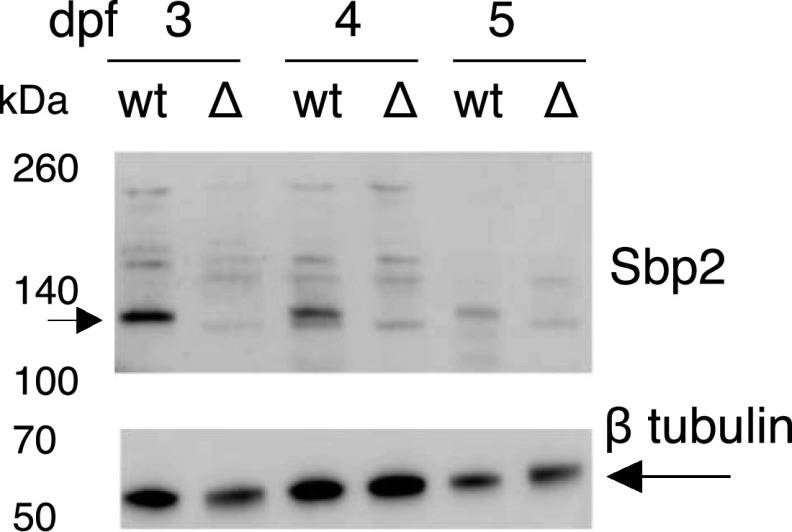
Figure 4.
Immunoblot analysis of Sbp2 expression.
Wild-type (wt) and sbp2 −/− (Δ) embryos at the indicated days post fertilization were lysed and two embryo equivalents were loaded onto a 4–12% gradient SDS–PAGE gel. Immunoblot was probed with a polyclonal antibody raised against human SBP2.
Acknowledgments
This image is the copyrighted work of the attributed author or publisher, and
ZFIN has permission only to display this image to its users.
Additional permissions should be obtained from the applicable author or publisher of the image.
Full text @ Life Sci Alliance