
|
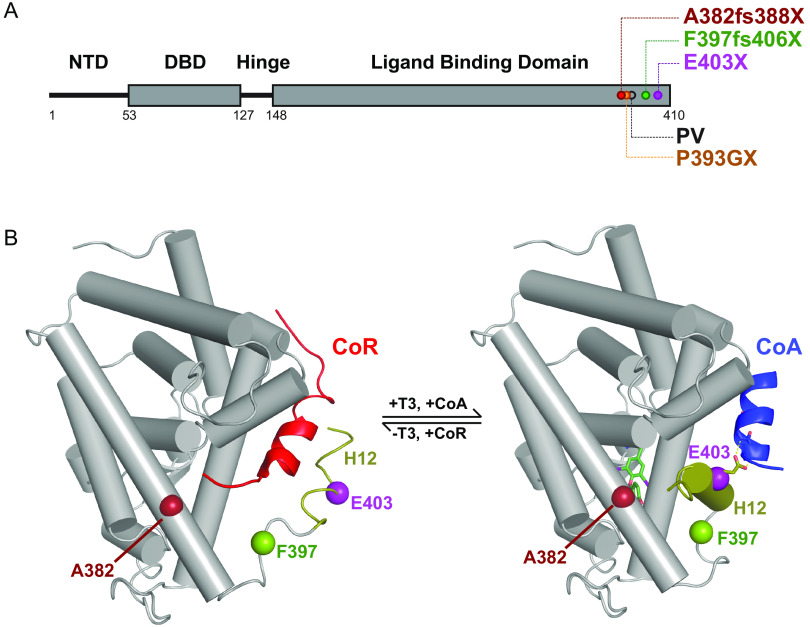
FIG 1 Location of human TRα mutations and WT TRα molecular switch. (A) Schematic representation of the full-length TRα, denoting the different functional domains of the receptor. Location of natural human mutations associated with RTHα (A382fs388X, F397fs406X, and E403X) and location of artificial mutation (P393GX) made for crystallization studies are indicated. (B) Structural model of TRα hormone-binding domain (PDB 2h79) in the unliganded state interacting with corepressor peptide (red) from the PPARα LBD:SMRT complex (PDB 1kkq) (left panel), and in the liganded state bound to coactivator peptide (blue) from the PPARα LBD:GRIP1 complex (PDB 1p54) (right panel). The position (A382, F397, E403) of natural human TRα mutations and the hydrogen bond between E403 and coactivator peptide are also shown.