
Figure 4
- ID
- ZDB-IMAGE-220302-198
- Genes
- Publication
- Trengove et al., 2022 - Functional Analysis of Zebrafish socs4a: Impacts on the Notochord and Sensory Function
- All Figures
- Figures for Trengove et al., 2022
|
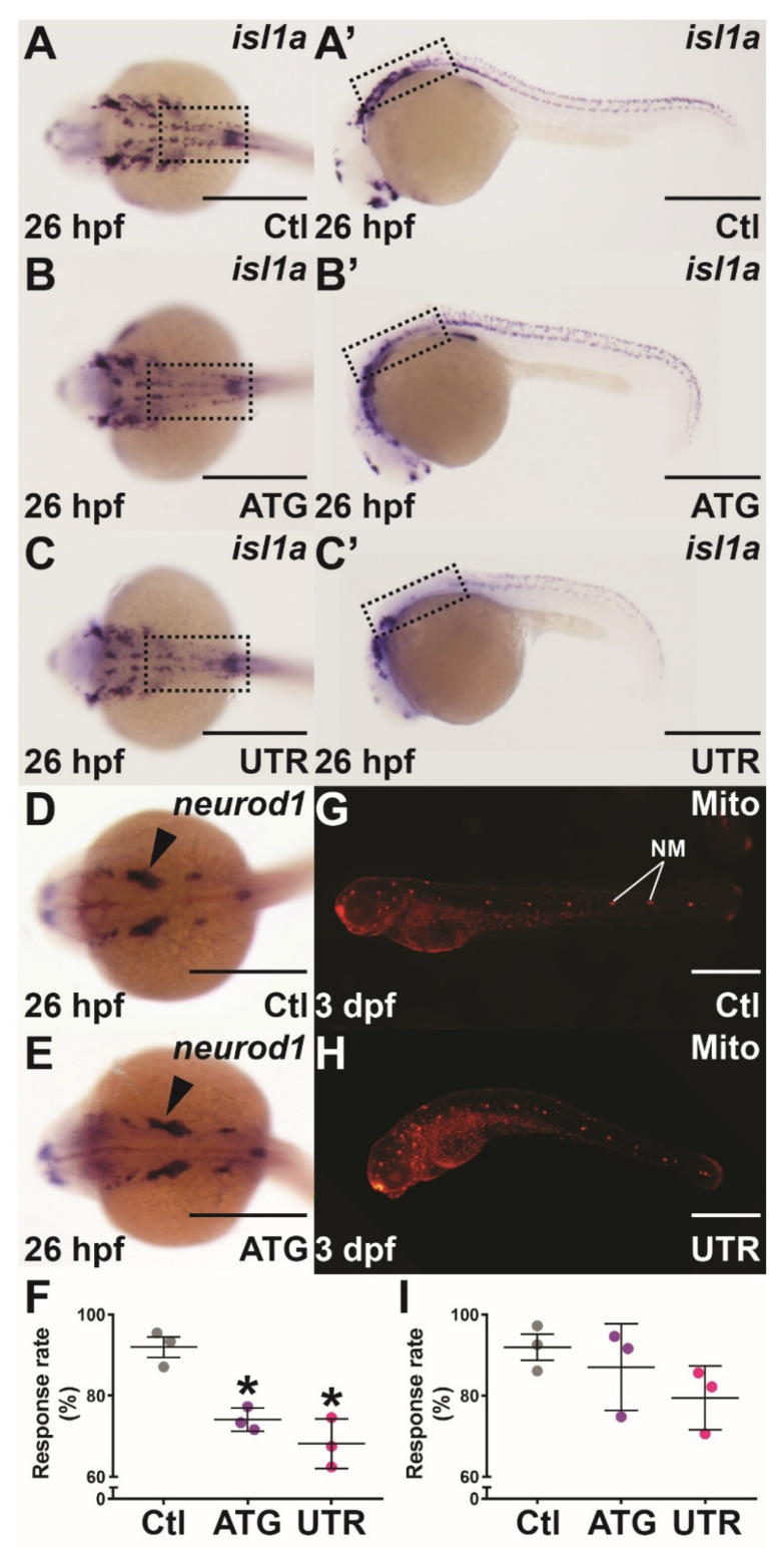
Figure 4 Mechanosensory analysis in socs4a knockdown embryos. Embryos injected with Ctl (A,A’,D,G), ATG (B,B’,E) or UTR (C,C’,H) MO were subjected to WISH with isl1a (A–C’) or neurod1 (D,E) at 26 hpf, or Mitotracker red (Mito) staining at 3 dpf (G,H) and imaged dorsally (A–E) and laterally (A’–C’,G,H), with the boxed areas highlighting the equivalent areas in the alternative images (A–C,A’–C’). Scale bars = 0.5 mm. Alternatively, the responsiveness to touch at 24 hpf (F) and water movement at 3 dpf (I) was quantified, with results presented for 3 independent experiments (n > 30 embryos in each group) along with mean ± SEM and statistical significance relative to Ctl (p < 0.05: *). Abbreviation: NM, neuromast.