
|
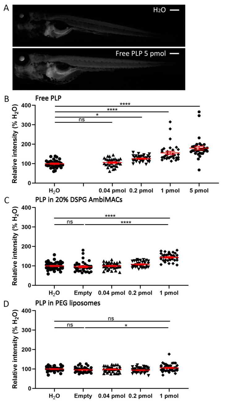
Figure 6
Liposome encapsulation of PLP decreased its effect on the systemic Gr transactivation activity. Embryos (3 dpf) of the Tg(GRE:GFP) line were injected with free or liposome-encapsulated PLP, and fluorescence microscopy images were taken at 24 hpi. (A) Representative images of Tg(GRE:GFP) embryos injected with H2O or 5 pmol PLP, showing the GFP signal, which is a readout for the transactivation activity of Gr, which increased after PLP injection. Scale bar = 200 μm. (B–C) The quantified GFP signals in the Tg(GRE:GFP) embryos at 24 hpi are shown after injection of different doses of free PLP (B), PLP encapsulated in AmbiMACs (20% DSPG) (C), and PLP encapsulated in PEGylated liposomes (D); H2O and empty liposome were injected as control. Statistical analysis was performed by one-way ANOVA with Bonferroni’s post hoc test. Significant increases in the GFP signal were observed when embryos were injected with 0.2–5 pmol free PLP and 1 pmol of PLP encapsulated in either AmbiMACs (20% DSPG) or in PEGylated liposomes. Each data point represents a single embryo, and the means ± SEM of the data accumulated from three independent experiments are shown in red. Statistically significant differences between groups are indicated by: ns, non-significant; * p < 0.05; **** p < 0.0001.