
Fig. 3.
- ID
- ZDB-IMAGE-220225-9
- Publication
- Diamond et al., 2022 - Computational anatomy and geometric shape analysis enables analysis of complex craniofacial phenotypes in zebrafish
- All Figures
- Figures for Diamond et al., 2022
|
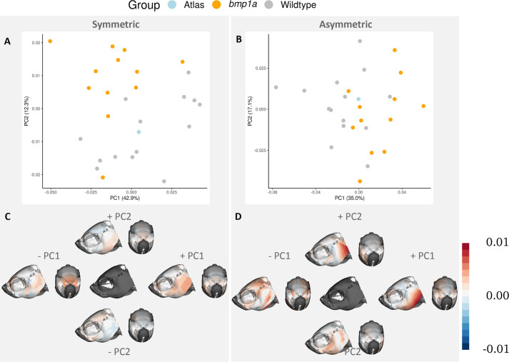
Fig. 3. First two principal components of symmetry analysis. PC plots show separation of groups (represented by color) along the first and second PCs (A,B). Heat maps of the same PCs represent where shape variation occurs across each axis (C,D). Columns represent symmetric (A,C) and asymmetric (B,D) components of shape variation. The central image in C and D represents mean shape of each component. Color in C and D represents the Procrustes distance between the average shape and the shape occupying the ends of each PC axis. Deeper colors represent larger differences, and the specific colors refer to differences in direction relative to the average image.