
|
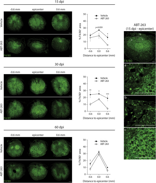
Fig. 6 ABT-263 treatment decreases the number of macrophages at the injury site Transversal sections at the lesion epicenter of an injured spinal cord at 15, 30 and 60 dpi treated with vehicle or ABT-263 and stained with the pan-macrophage marker F4/80 (green). Scale bars, 200 μm. The area of F4/80+ tissue was measured at the lesion epicenter and 600 μm rostrally and caudally from the epicenter. The panel on the right displays zoomed images of three different zones (a–c) of an ABT-263-treated spinal cord transversal section stained with F4/80 at 15 dpi. Macrophages form a network inside the central lesion core, but they can be individually distinguished outside of it. Scale bars, 100 μm. Measurements are expressed as a percentage of the total cross-sectional area. n (15 dpi) = 3–4; n (30 dpi) = 3–4; n (60 dpi) = 2–3. Data are presented as mean ± SEM. ∗p < 0.05, ∗∗p < 0.01, ∗∗∗p < 0.001, ABT-263 versus vehicle.