
|
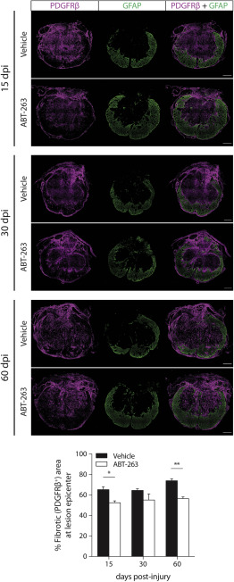
Fig. 5 Targeting senescent cells leads to a reduction of the fibrotic scar Transversal sections at the lesion epicenter of an injured spinal cord at 15, 30, and 60 dpi treated with vehicle or ABT-263 and stained with the fibrotic scar marker PDGFRβ+ (magenta) and with the astrocytic scar marker GFAP (green). The fibrotic scar area was evaluated by normalizing the PDGFRβ+ area to the total cross-sectional area at the lesion epicenter. GFAP+ tissue surrounds the fibrotic core. Scale bars, 200 μm. n (15 dpi) = 3–4; n (30 dpi) = 3–4; n (60 dpi) = 2–3. The lower panel shows the percentage of fibrotic tissue in the injury core as quantified at 15, 30 and 60 dpi. Data are presented as mean ± SEM. ∗p < 0.05, ∗∗p < 0.01, ABT-263 versus vehicle.