
|
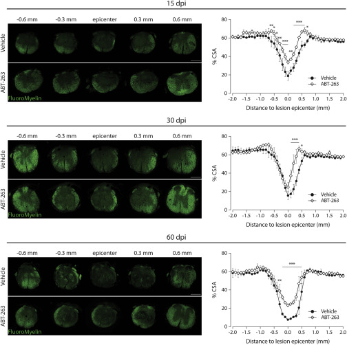
Fig. 4 White matter sparing is increased after targeting senescent cells with ABT-263 Transversal sections at different distances from the lesion epicenter of an injured spinal cord at 15, 30 and 60 dpi, treated with vehicle or ABT-263, and stained with FluoroMyelin (green) and the corresponding quantifications. White matter sparing was assessed by normalizing the area stained with FluoroMyelin (green) to the total cross-sectional area (CSA) of spinal cord sections every 100 μm ranging from 2 mm rostral and 2 mm caudal to the lesion epicenter. Scale bars, 500 μm. n (15 dpi) = 3–4; n (30 dpi) = 3–4; n (60 dpi) = 2–3. Data are expressed as % CSA and presented as mean ± SEM. ∗p < 0.05, ∗∗p < 0.01, ∗∗∗p < 0.001, ABT-263 versus vehicle.