IMAGE

Fig. 3

ID
ZDB-IMAGE-220219-17
Source
Figures for Zhang et al., 2022
Image
Figure Caption

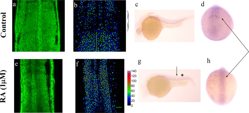
Fig. 3 Somitogenesis at high Erk activity.

ad Control: normal development. a Erk activity in DREKA embryos at 14 somite stage: dark nuclei point to high Erk activity. b The false color images represent the Erk activity averaged over a 30 μm perpendicular stack of images. c ISH against xirp2a (a marker of somite boundaries) in 30 hpf embryos. d IHC against Mesp2a, a marker of the last somite boundary at 14 somites (arrow). eh Somitogenetic development in DREKA embryos incubated from one-cell stage in 10 μM DEAB in presence of 1 μM RA from the ten somite stage (arrow in g). e, f The Erk activity is high throughout the PSM. g ISH against xirp2a in 30hpf embryos. Somitogenesis is impaired past 13 somites (indicated by a *) with unclear somite boundaries. h IHC against Mesp2a show that it is not expressed past 13 somite stage (arrows in d and h). Scale bar: 50 μm.

Acknowledgments
This image is the copyrighted work of the attributed author or publisher, and ZFIN has permission only to display this image to its users. Additional permissions should be obtained from the applicable author or publisher of the image. Full text @ Commun Biol