
|
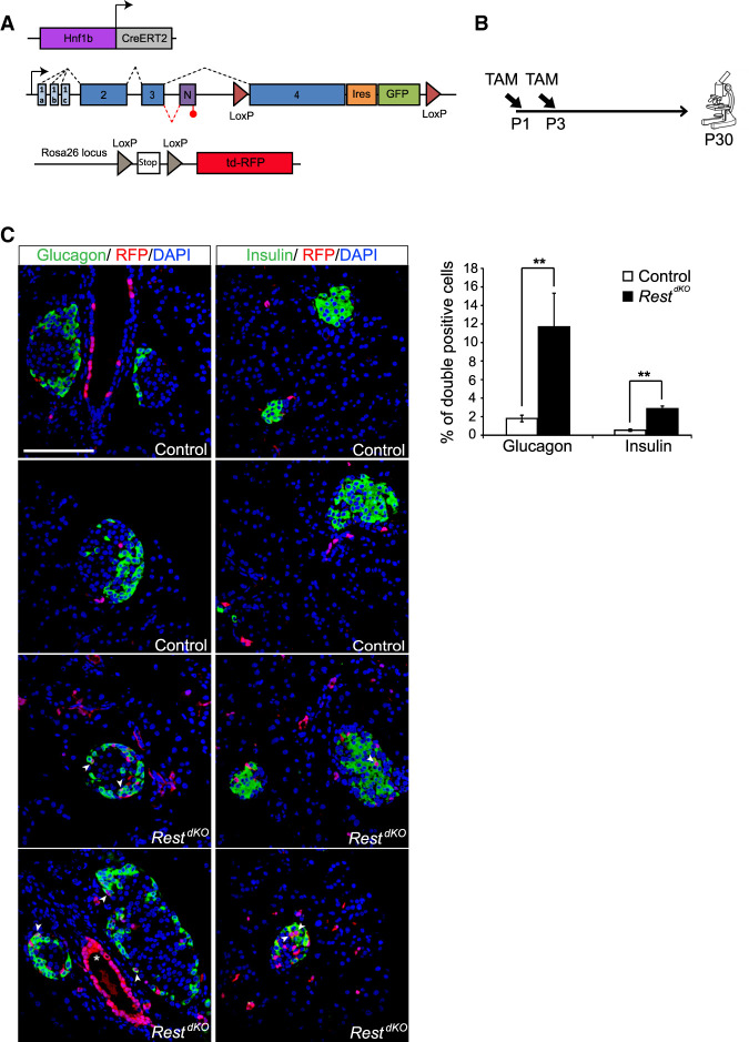
Figure 4.
Pancreas-specific inactivation of Rest in neonatal ducts. (A) Schematic of genetic models used to inactivate Rest and activate RFP expression in duct cells and progeny. Hnf1b-CreERT2 is a BAC transgenic that specifically marks duct cells (Solar et al. 2009), as well as non-Rest-expressing ∂ cells in reporters that are excised with high efficiency (Rovira et al. 2021), but not other endocrine or acinar cells. (B) Schematic of the lineage tracing experiment. Tamoxifen was given to mothers at day 1 (P1) and day 3 (P3) after delivery, and mice were analyzed at P30. Hnf1b-CreERT2;Rosa26RFP control mice were also treated. (C) Representative images of double-positive RFP (red) and insulin (green) cells and of double-positive RFP (red) and glucagon (green) cells in RestdKO and control mice. The graph shows RFP-expressing glucagon and insulin cells in RestdKO versus control mice. n = 5–6 mice per each group. Arrowheads indicate double-positive cells, and an asterisk marks examples of cells in a duct, which were very efficiently labelled. Scale bar, 100 µm. Error bars are SEM. Student's t-test; (**) P < 0.01.