
|
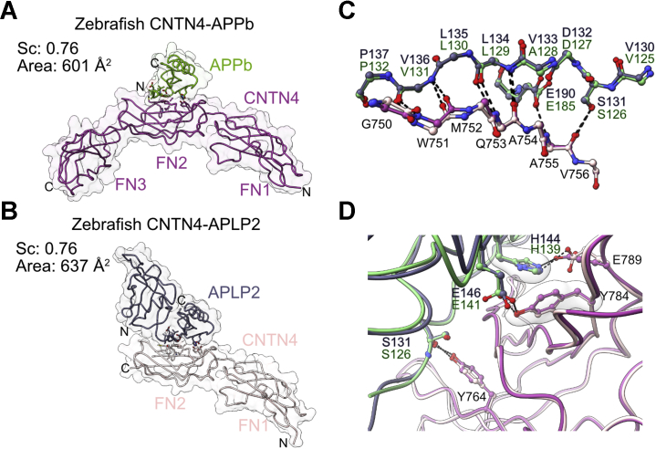
Figure 6
Crystal structures of zebrafish CNTN4 bound to APPb and APLP2.A, crystal structure of the copper-binding domain of zebrafish APPb in complex with CNTN4(FN1–FN3). B, crystal structure of the fusion protein between zebrafish CNTN4(FN1–FN2) and the E1 domain of APLP2. In A and B, the structures are shown in coil and surface representations, whereas the letters N and C indicate the N and C termini, respectively. C, detailed view of side chain–side chain interactions at the CNTN4–APPb and CNTN4–APLP2 interfaces. Dashed lines indicate potential hydrogen bonds or salt bridges, whereas translucent surfaces highlight residues involved in packing interactions. D, main-chain hydrogen bonding network, indicated by dashed lines, between antiparallel β-strands in CNTN4 and APPb–APLP2. The structures were superimposed using the copper-binding domains of APPb and APLP2 (RMSD of 0.57 Å over 62 Cα pairs). APLP2, amyloid beta precursor like protein 2; APP, amyloid precursor protein; CNTN4, contactin 4; FN1, first FN repeat; FN2, second FN repeat; FN3, third FN repeat.