
Fig. 8
- ID
- ZDB-IMAGE-220124-3
- Antibodies
- Publication
- Taler et al., 2019 - Lysyl hydroxylase 3 is required for normal lens capsule formation and maintenance of lens epithelium integrity and fate
- All Figures
- Figures for Taler et al., 2019
|
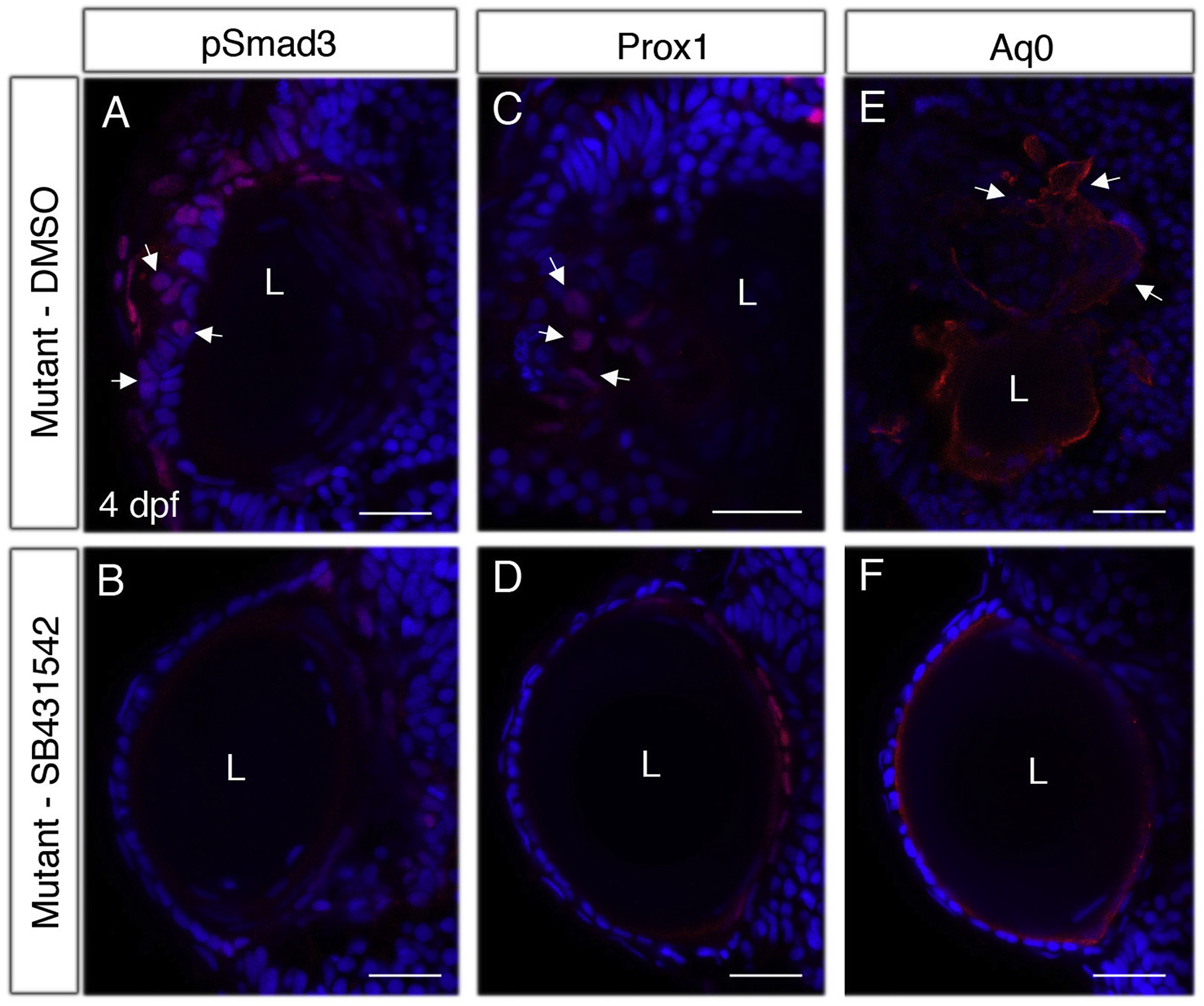
Fig. 8 Inhibition of TGFβ signaling restores normal expression of pSmad3, Prox1 and Aquaporin 0. (A–F) Confocal images of sections from eyes of 4 dpf plod3vu222/vu222 embryos treated with vehicle (DMSO) (A,C,E) or with SB-431542 (B,D,F) to inhibit TGFβ signaling via TGF type I receptor. Sections were labeled for pSmad3 (red, A,B), Prox1 (red, C,D) and Aq0 (red, E,F). In all panels, nuclei are labeled with DAPI (blue). Arrows in A,C,E point at abnormal expression of pSmad3, Prox1 and Aq0, respectively, in the cell masses. Data is representative of the following number of eyes: A = 9, B = 11, C = 6, D = 12, E = 7, F = 10. L, lens. Scale bars are 20 μm.
Reprinted from Developmental Biology, 458(2), Taler, K., Weiss, O., Rotem, S., Rubinstein, A.M., Seritrakul, P., Gross, J.M., Inbal, A., Lysyl hydroxylase 3 is required for normal lens capsule formation and maintenance of lens epithelium integrity and fate, 177-188, Copyright (2019) with permission from Elsevier. Full text @ Dev. Biol.