
Fig. 1
- ID
- ZDB-IMAGE-220107-27
- Publication
- Park et al., 2021 - Flavin Adenine Dinucleotide Depletion Caused by electron transfer flavoprotein subunit alpha Haploinsufficiency Leads to Hepatic Steatosis and Injury in Zebrafish
- All Figures
- Figures for Park et al., 2021
|
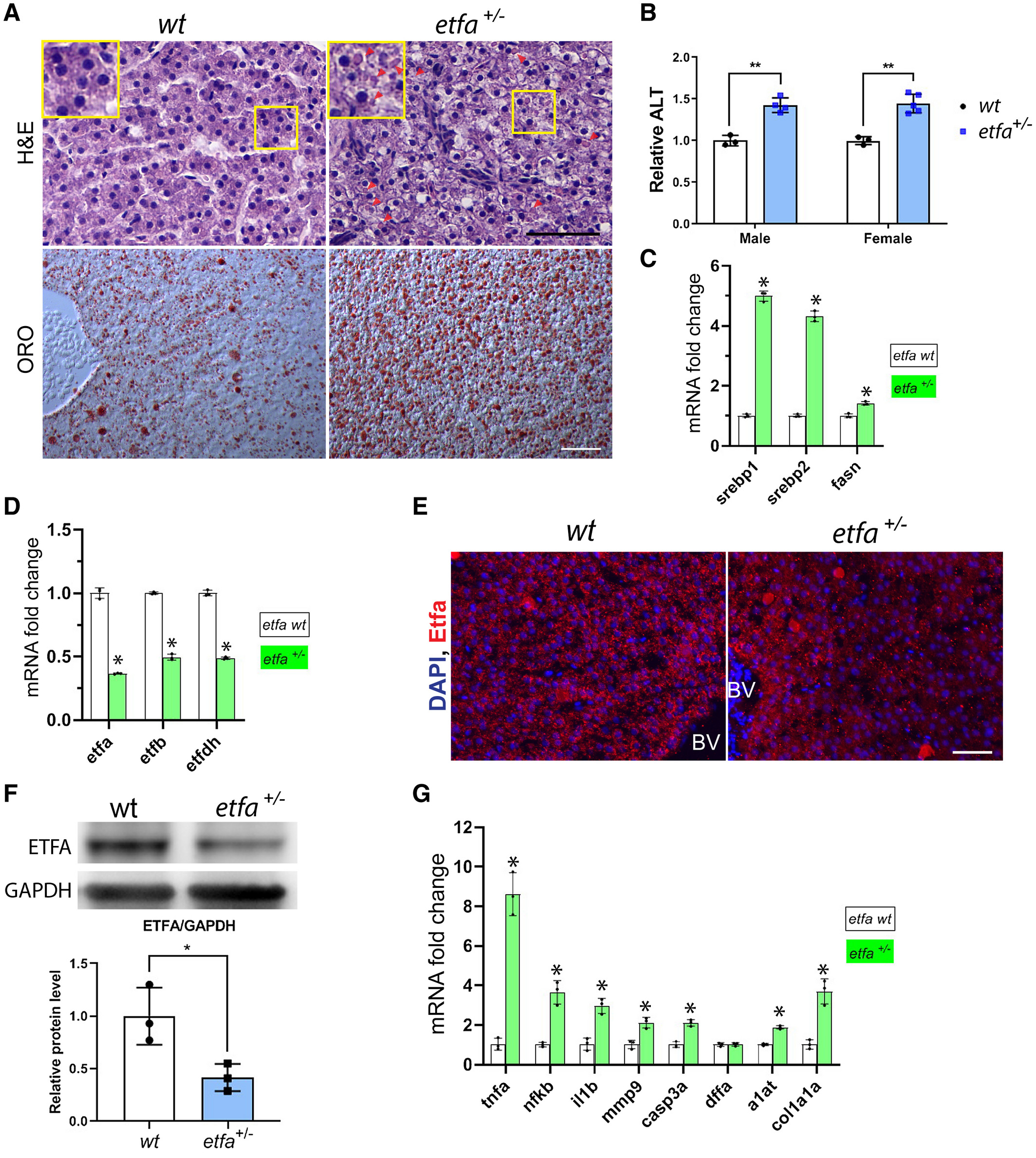
Fig. 1 Etfa haploinsufficiency induces hepatic injury and steatosis in adult zebrafish. (A) Representative images show hepatocyte ballooning and accumulation of cytosolic components (note eosinophilic cytoplasmic droplets marked with arrow heads) in hepatocytes in etfa+/− livers (H&E staining). Magnified views (yellow rectangles) are shown. In the bottom panels, sections were stained with ORO (representative images are shown, n = 5/5); scale bar, 50 µm. (B) Relative serum ALT levels in etfa+/− siblings compared to the wild type (n = 3 to 5). (C) Livers were harvested, and pooled total RNA was subjected to qRT-PCR (see Materials and Methods) to determine relative mRNA expression genes associated with de novo lipogenesis (srebp1, srebp2, and fasn). (D) Relative mRNA expression of genes of etfa, etfb, and etfdh (*P < 0.05 vs. the wild type). (E) Immunofluorescence staining with anti-Etfa antibody (red) on wild-type and etfa+/− liver sections; scale bar, 50 µm. Nuclei were stained with DAPI. (F) Relative amount of Etfa protein in wild-type and etfa+/– liver lysates (n = 3). (G) Relative mRNA expression of genes associated with inflammation (tnfa, nfkb, il1b, and mmp9), cell death (casp3a and dffa), injury (a1at), and fibrosis (col1a1a). Pooled RNAs (from three livers per group) were used for qRT-PCR analysis. *P < 0.05, **P < 0.005 vs. the wild type. Data in bar graphs represent mean ± SD. Abbreviations: BV, blood vessel; DAPI, 4′,6-diamidino-2-phenylindole; GAPDH, glyceraldehyde 3-phosphate dehydrogenase.