
|
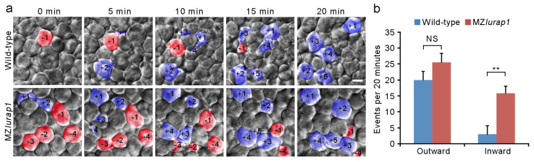
Figure 4 Defective radial intercalation of DEL cells in MZlurap1 mutant embryos. (a) Still frames from representative time-lapse imaging of DEL cell movements in the animal pole region of wild-type and MZlurap1 mutant embryos at 50% epiboly. The focal plane was set at the outermost layer of the DEL immediately underlying the EVL. Cells undergoing outward movements are fake color labeled blue, and are attributed with positive numbers, while cells undergoing inward movements are labeled red, and are attributed with negative numbers. Scale bar: 10 μm. (b) Statistical analysis compares inward and outward movements in the same focal plane during a period of 20 min. The results were analyzed independently in a blended manner. Data were collected from 5 embryos for each group in three independent experiments. Bars represent the mean value ± s.d. (NS, not significant; **, p < 0.01).