IMAGE

Figure 8

ID
ZDB-IMAGE-211216-155
Source
Figures for Dalum et al., 2021
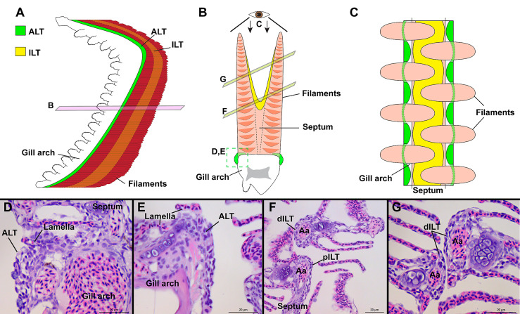
Image
Figure Caption

Figure 8

General organization of the structure lymphoid tissues in zebrafish gills. Schematic representations of the localization of the ALT (green) and ILT (yellow) within adult zebrafish gills. The ALT runs along the base of filaments while the ILT is found on the inner (afferent) aspect of filaments, on top of the interbranchial septum. (A) illustrates a gill arch as observed from the side. (B) represents a transversal plane through the gill arch, as illustrated on (A). (C) shows a gill arch as observed from above the filaments, as illustrated in (B). (D, E) Paraffin sections (2 µm) displaying the ALT, as illustrated in (B). (F, G) Paraffin section (2 µm) displaying the proximal ILT (pILT) and distal ILT (dILT), as illustrated in (B). Aa, Afferent artery; ALT, Amphibranchial Lymphoid Tissue; ILT, Interbranchial Lymphoid Tissue; dILT, distal Interbranchial Lymphoid Tissue and pILT, proximal Interbranchial Lymphoid Tissue. Scale bars: 20 µm (D–G).

Acknowledgments
This image is the copyrighted work of the attributed author or publisher, and ZFIN has permission only to display this image to its users. Additional permissions should be obtained from the applicable author or publisher of the image. Full text @ Front Immunol