
|
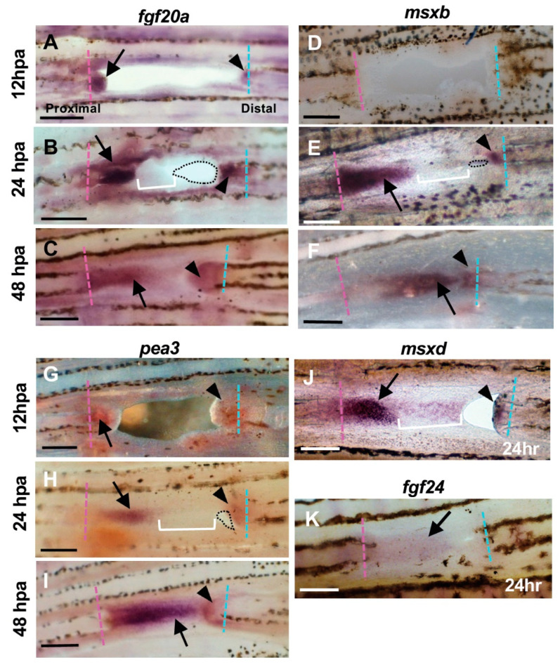
Figure 3
Expression of regeneration-related genes during hole closure. (A–C) fgf20a expression. (A) At 12 h post-amputation (hpa). The fgf20a expression was observed in the cells of the proximal margin (arrow), and distal margin (arrowhead) of the hole. (B) At 24 hpa. In the proximal margin, fgf20a was strongly expressed in the cells of the inner half of the regenerated tissue (arrow), whereas the expression was faint in the apical half of the tissue (bracket). In the distal margin, fgf20a expression was also observed, but the area of expression was small (arrowhead). Dotted lines show the remaining hole. (C) At 48 hpa. The fgf20a expression was maintained in both proximal- and distal-derived tissues (arrow and arrowhead, respectively), and both domains were close together. (D–F) msxb expression. (D) At 12 hpa, msxb was not expressed. (E) At 24 hpa, in the proximal margin, msxb was strongly expressed in the cells near the cut surface of the regenerated tissue (arrow). The expression was faint in the distal half of the tissue that filled the hole (bracket). In the distal margin, msxb expression was restricted to a small area (arrowheads). Dotted lines show the remaining hole. (F) At 48 hpa, msxb was maintained in both proximal- and distal-derived tissues (arrow and arrowhead, respectively). (G–I) pea3 expression. (G) At 12 hpa, pea3 expression was observed in the cells both in the proximal and distal margins of the hole (arrow and arrowhead, respectively). (H) At 24 hpa, pea3 was expressed in the cells of the inner half of the regenerated tissue in the proximal margin (arrow), whereas the expression was weakened in the apical half of the tissue (brackets). In the distal margin, pea3 expression was also observed, although the expression domain was small (arrowhead). Dotted lines show the remaining hole. (I) At 48 hpa, pea3 expression was maintained in both proximal- and distal-derived tissues (arrow and arrowhead, respectively). (J) msxd expression at 24 hpa. In the proximal margin, msxd was strongly expressed in the cells of the inner half of the regenerated tissue (arrow), whereas the expression was weakened in the apical half of the tissue (bracket). In the distal margin, msxd expression was also observed, but the area of expression was restricted (arrowhead). (K) fgf24 expression at 24 hpa. The fgf24 was weakly expressed in the epithelium of the regenerated tissue from the proximal margin (arrow). Expression in the distal margin was unclear. Scale bars = 300 μm.