
|
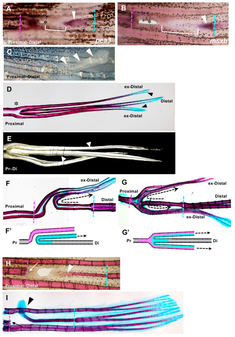
Figure 6
Fin ray regeneration of fin ray from the distal margin of the hole. (A,B) Strong expression of both msxb and pea3 was observed at both the proximal and distal sides. Arrowheads indicate gene expression that was extended from the distal side to the proximal side. In addition, a distal margin-derived sheet-like tissue was observed (yellow brackets). (C) Seven days after excision, a rod-like lateral protrusion was formed from the position of the hole closure (arrowheads). (D) An example of the skeletal pattern that was fully regenerated from a hole-excised caudal fin. The original and regenerated fin rays are shown. Regenerated fin rays laterally protruded from the position of the hole with extra branching (asterisk). The distal ends of the regenerated fin rays (indicated as ex-distal) reached the same length as the original fin ray and were stained with alcian blue (arrowheads). (E) A 3D reconstruction of the regenerated extra fin ray shown in (D) using the COMBI method. Bifurcation of the regenerated fin ray was observed (arrows). (F,G) Examples of the skeletal pattern at the branching position of the hole-excised caudal fin. The direction of the regenerated fin ray extending from the distal margin is indicated with dotted arrows. (F’) and (G’) are schema that represent the fin ray patterns shown in (F,G), respectively. In the schema, fin rays regenerated from the distal margin are indicated in blue, and those regenerated from the proximal margin are indicated in pink. In (F) the fin ray that regenerated from the distal margin protruded to either side of the fin at the position where it collided with the fin ray that regenerated from the proximal margin, and then bent distally (n = 3/9). In (G) a hemi-ray of the distal-derived fin ray and that of the proximal-derived fin ray merged to form one fin ray and protruded to both sides of the fin (n = 6/9). (H,I) A fin ray regenerated from a distal margin after prolonged and repeated excision of tissue at the proximal margin of the hole. (H) After one week, the regenerated fin ray was formed in the same plane of the original fin rays and as a continuous structure of the proximal edge of the original fin ray (arrowhead, n = 3). (I After 2 weeks of regeneration, the distal end of the regenerated fin ray protruded laterally (arrowhead, n = 3). The distal end was stained with alcian blue (arrowhead), as was the original fin ray. During the regeneration process from the distal cut edge, regeneration from the proximal margin did not occur because of repeated excision (arrows).