
Image
|
Figure Caption
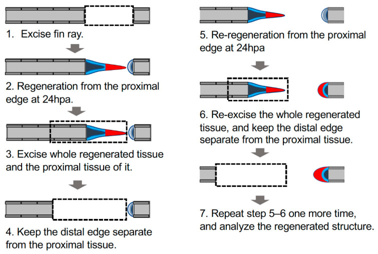
Figure 5
Schema of manipulation to investigate autonomous sheet-like tissue formation in the distal margin. Schema of the experimental procedure; the detailed procedure is described in the Materials and Methods section. Dashed rectangles indicate excision sites of the fin ray; gray, fin ray; light blue, epithelium of the cut surface; dark blue, mesenchyme of the cut surface; red, the sheet-like tissue formed on the proximal margin (triangle) or an equivalent structure on the distal margin (crescent shape).
Acknowledgments
This image is the copyrighted work of the attributed author or publisher, and
ZFIN has permission only to display this image to its users.
Additional permissions should be obtained from the applicable author or publisher of the image.
Full text @ J Dev Biol