
|
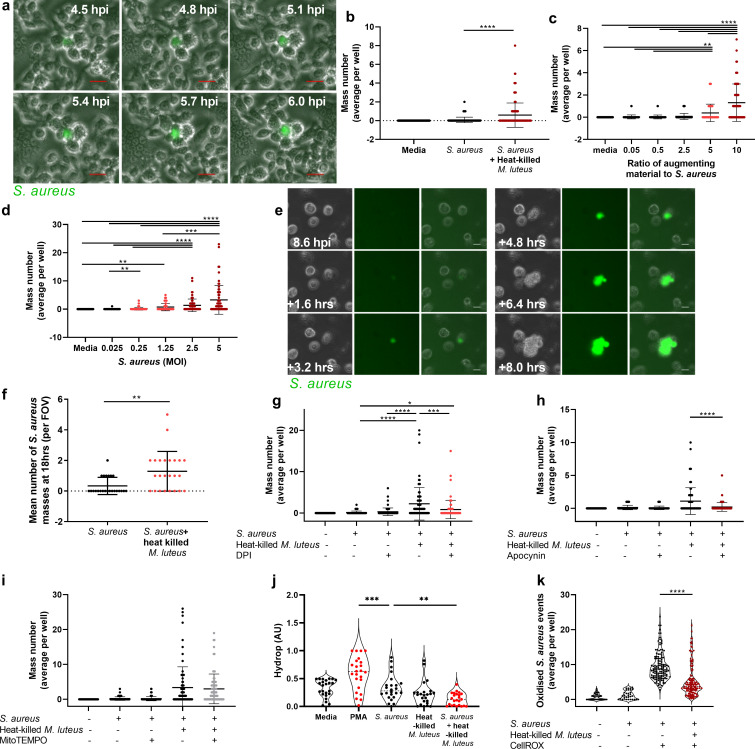
Fig 5
A Images of GFP-S. aureus mass formation within RAW264.7 cells, scale 20 μm B RAW264.7 cells infected with GFP S. aureus (MOI 5) with or without heat-killed M. luteus (MOI 50), (n = 4), ****p<0.0001 C RAW264.7 cells infected with GFP S. aureus (MOI 5) with or without heat-killed M. luteus (ratio to S. aureus, 10, 5, 2.5, 0.5 or media control), (n = 4), **p<0.008; ****p<0.0001 D RAW264.7 cells infected with GFP S. aureus (MOI 5, 2.5, 1.25, 0.25, 0.025 or media control) with or without of heat-killed M. luteus (MOI 50), (n = 4), **p<0.003; ***p<0.0008; ****p<0.0001 E-F MDMs infected with GFP S. aureus (MOI 5) with or without heat-killed M. luteus (MOI 50) E images of GFP S. aureus mass formation within human MDMs, scale 20 μm F number of S. aureus masses observed (n = 3), **p<0.003 G RAW264.7 cells infected with GFP S. aureus (MOI 5) in the presence or absence of heat-killed M. luteus (MOI 50), either with or without DPI (2 μM), (n = 4), *p<0.05;***p<0.0004; ****p<0.0001 H RAW264.7 cells infected with GFP S. aureus (MOI 5) in the presence or absence of heat-killed M. luteus (MOI 50), either with or without apocynin (500 μM), (n = 4), ****p<0.0001 I RAW264.7 cells infected with GFP S. aureus (MOI 5) in the presence or absence of heat-killed M. luteus (MOI 50), either with or without mitoTEMPO (1 μM), (n = 4, non-significant) J RAW264.7 cells infected with GFP S. aureus (MOI 5) with or without heat-killed M. luteus (MOI 50) with Hydrop used to visualise hydrogen peroxide (n = 4, violin plot with median values shown), **p<0.007; ***p<0.0004 K RAW264.7 cells infected with CellROX-stained GFP S. aureus (MOI 50) to visualise intracellular oxidation in the presence or absence of heat-killed M. luteus (MOI 50), (n = 4, violin plot with median values shown), ****p<0.0001. In panels B, F, H, I and K, a two-tailed Mann Whitney test was used, in panels C, D, G, and J, a Kruskal-Wallis test with Dunn’s post hoc test was used. Where used, error bars show mean +/- SD.