
Image
|
Figure Caption
Figure 3
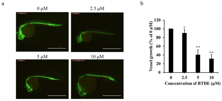
BTDE blocks intersegmental vessel formation in zebrafish embryos. (a) Lateral view of Tg (flk1: EGFP) zebrafish embryos at 16 hpf. Embryos were treated with different concentrations of BTDE. Vessels of zebrafish embryos were observed using a fluorescence microscope, photos were recorded by inverted fluorescence microscope (scale bar: 1.2 mm). (b) Quantification of intersegmental vessel growth induced by BTDE. Values represent the means ± SD of three independent experiments. * p < 0.05, ** p < 0.01 versus medium control.
Figure Data
Acknowledgments
This image is the copyrighted work of the attributed author or publisher, and
ZFIN has permission only to display this image to its users.
Additional permissions should be obtained from the applicable author or publisher of the image.
Full text @ Mar. Drugs