
|
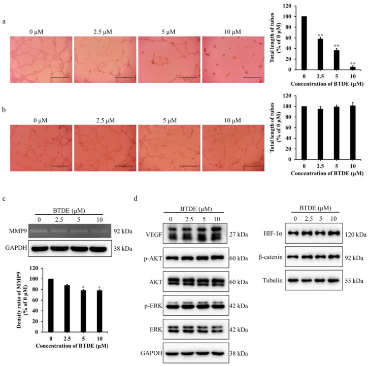
Figure 2
BTDE reduces HUVECs tube formation and MMP9 activity. (a) HUVECs was pretreated with BTDE for 24 h, then seeded on matrigel for 20 h, capillary-like structures of HUVECs were recorded by inverted microscope (original magnification, 4×; scale bar: 600 μm) and total length of tubes was measured by Image J software. (b) Different concentrations of BTDE were added after tubes have established on matrigel for 8 h, and incubated for another 6 h. Tubular structures were observed by inverted microscope (original magnification, 4×; scale bar: 600 μm) and total length of tubes compared with 0 μM was measured by Image J software. (c) Gelatin zymography experiment was used to detect the MMP9 activity of HUVECs after 24 h treatment of BTDE, GAPDH was used as an internal control. (d) Western blot was used to measure the VEGF, HIF-1α, β-catenin, AKT, and ERK as well as their phosphorylation levels in HUVECs treated with BTDE for 24 h. Data represent mean ± SD of three independent experiments. * p < 0.05, ** p < 0.01 versus control.