
Figure 8
- ID
- ZDB-IMAGE-211201-207
- Genes
- Publication
- Schmitner et al., 2021 - Differential Responses of Neural Retina Progenitor Populations to Chronic Hyperglycemia
- All Figures
- Figures for Schmitner et al., 2021
|
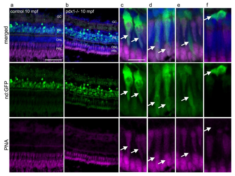
Figure 8
neurod:GFP expressing progenitors give rise to cones and rods in pdx−/− diabetic zebrafish. Expression of GFP in tg(neurod:GFP) in the INL in control fish (a) and in the INL and in the ONL in pdx1−/− fish (b). (c–f) Higher magnification views of photoreceptors from pdx1−/− samples as in (b). GFP expression overlaps with PNA in cells with cone morphology (c,d, arrows), while rod-shaped cells (e, arrow) are PNA negative. Compact cells located basally in the ONL, presumed to be undifferentiated, do not label with PNA (f, arrow). Sections were counterstained with DAPI (blue). Scale bar: 50µm in a and b and 10µm in (c–f). (INL, inner nuclear layer; ONL, outer nuclear layer; PRL, photoreceptor layer).