
Figure 5
- ID
- ZDB-IMAGE-211201-196
- Genes
- Publication
- Raby et al., 2021 - Loss of Polycomb Repressive Complex 2 Function Alters Digestive Organ Homeostasis and Neuronal Differentiation in Zebrafish
- All Figures
- Figures for Raby et al., 2021
|
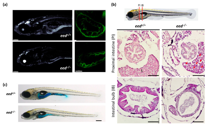
Figure 5
Structure of the intestine at 9–11 dpf: (a). confocal imaging of the anterior part (left, scale bar is 150 µm) and of the intestine bulb (right, scale bar is 50 µm) of transgenic Tg (actb2:GFP-Hsa.UTRN)e116 zebrafish larvae, wild-type (up) or lacking eed function (down) at 9 dpf; (b) histological sections stained with hematoxylin and eosin at the levels of the proximal intestine (PI) and the intestinal bulb (IB) as indicated from eed+/+ (left) and eed−/− (right) siblings at 11 dpf. Red arrows show macrovesicles. L, liver. Scale bar is 50 µm; (c) Smurf assay performed on eed+/+ and eed−/− siblings at 11 dpf. Scale bar is 200 µm.