
Fig. 4
- ID
- ZDB-IMAGE-211116-4
- Publication
- Ghaddar et al., 2021 - Deleterious Effects of Overfeeding on Brain Homeostasis and Plasticity in Adult Zebrafish
- All Figures
- Figures for Ghaddar et al., 2021
|
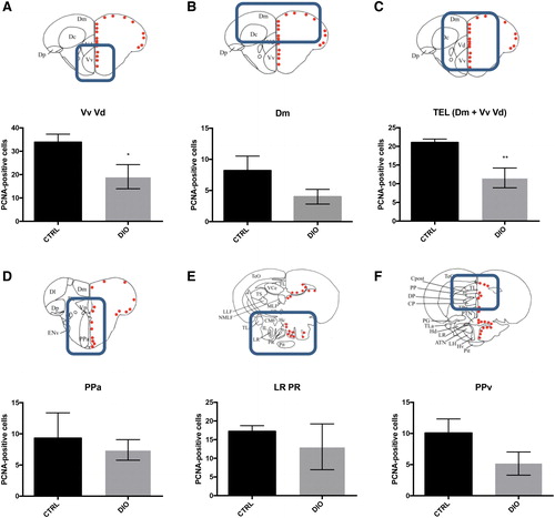
Fig. 4 DIO impairs neurogenesis in the forebrain of adult zebrafish. (A–F) Statistical analysis of the number of PCNA-positive cells in CTRL and DIO zebrafish. A significant decrease in proliferative activity was observed between CTRL and DIO group in the Vv-Vd and in the telencephalic region (Dm + Vv-Vd). Only a decreasing trend was observed in the Dm, PPa, PPv, and in the region surrounding the LR PR regions (n = 6). n = number of brains studied. Bar graph: SEM. Student's t-test: *p < 0.05; **p < 0.01. Scale bar = 32 μm. The brain schemes correspond to the transversal sections of the zebrafish brain for the region studied and showing the main brain domains/nuclei according to the Zebrafish Brain Atlas from Wullimann et al. and were adapted from Menuet et al.84,85 Dm, medial zone of dorsal telencephalic area; LR, lateral recess of diencephalic nucleus; PCNA, Proliferating Cell Nuclear Antigen; PPa, parvocellular preoptic nucleus, anterior part; PPv, periventricular pretectal nucleus; PR, posterior recess of diencephalic ventricle. Color images are available online.