
|
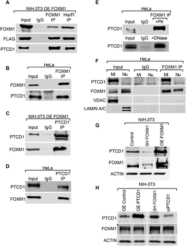
Fig. 5 FOXM1 and PTCD1 are protein binding partners. (A) PTCD1 in FOXM1 immunoprecipitates. NIH-3T3 cells expressing FOXM1 (OE FOXM1), which contains HIS and FLAG tags, were subjected to 2-step affinity chromatography using anti-FLAG and anti-HIS affinity columns. The elutes were analyzed by Western blot using antibodies for FOXM1, FLAG, and PTCD1. Cells incubated with IgG were used as controls. (B) Immunoblots show immunoprecipitation (IP) of endogenous FOXM1 with PTCD1 in HeLa cells. (C, D) FOXM1 in PTCD1 immunoprecipitates. (C) NIH-3T3 cells expressing FOXM1 (OE FOXM1) or (D) HeLa cells immunoprecipitated using PTCD1 antibody. Immunoblots show IP of endogenous PTCD1 protein with FOXM1. (E) Proteinase K and DNase treatments do not influence interactions of endogenous FOXM1 with PTCD1. Total wild-type HeLa cell lysates were incubated with 10 µg/ml proteinase K (PK) or DNase for 30 min. FOXM1 IP experiments were performed, and blots were probed with PTCD1 antibodies. (F) FOXM1 and PTCD1 are bound in mitochondrial lysates. FOXM1 or nonspecific isotype-matched IgG was incubated with purified mitochondrial (Mi) and nuclear (Nu) fractions from HeLa cells. Immunoprecipitates were resolved on SDS–PAGE and probed for PTCD1, FOXM1, VDAC (mitochondrial protein), and LAMIN A/C (nuclear protein). (G) FOXM1 directly correlates with PTCD1 protein levels. Immunoblots show FOXM1 and PTCD1 protein levels in total cell lysates of NIH-3T3 cells that stably express control, shFOXM1, and OE FOXM1. ACTIN was used as a loading control. (H) Changes in PTCD1 does not affect FOXM1 levels. NIH-3T3 cells were transiently transfected with the indicated constructs. Immunoblots show FOXM1 and PTCD1 protein levels in total lysates of NIH-3T3 cells expressing OE CONTROL, OE PTCD1, siCONTROL, and siPTCD1. β -ACTIN was used as a loading control.