
|
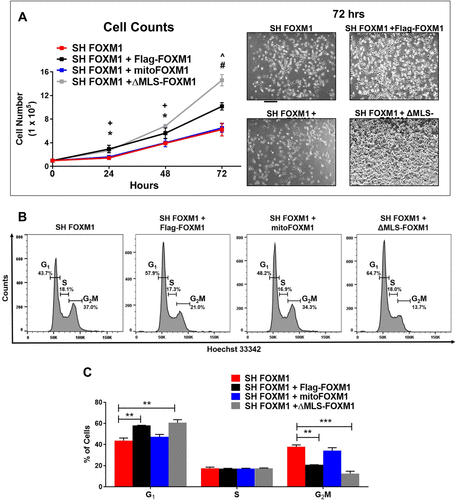
Fig. 4 Translocation of FOXM1 to the mitochondria does not affect proliferation. (A) Growth curves of SH FOXM1 cells transfected with FLAG-FOXM1, mitoFOXM1, or ΔMLS-FOXM1 constructs. Cells were counted at 24, 48, and 72 h. Representative images are shown for each group at 72 h. Magnification: ×5; 100 μm scale bar. Graph represents data from n = 5 replicates presented as mean ± SEM, n = 5, ANOVA, Bonferroni’s post test. * indicates statistical significance of p < 0.05 comparing SH FOXM1 + Flag-FOXM1 to SH FOXM1. + indicates statistical significance of p < 0.05 comparing SH FOXM1 + ΔMLS-FOXM1 to SH FOXM1. #indicates statistical significance of p < 0.001 comparing SH FOXM1 + ΔMLS-FOXM1 to SH FOXM1. ^ indicates statistical significance of p < 0.001 comparing SH FOXM1 + Flag-FOXM1 to SH FOXM1. (B) Effects of FOXM1 localization on cell cycle distribution. SH FOXM1 cells were transfected with the indicated MLS mutant constructs, and cell cycle analysis was measured using Hoechst 3342 and flow cytometry 72 h post–serum starvation. (C) Representative quantitative chart depicts cell cycle phase. Graph represents G1, S, and G2/M phases and values are presented as mean ± SEM from four replicates. Red dotted line represents OCR values from NIH-3T3 cells stably expressing SH FOXM1 alone. Statistical significance is indicated with an asterisk (*); *p < 0.05, **p < 0.01, and ***p < 0.001.