
|
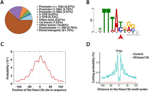
Fig. 3 Hoxa13b-binding sites and motif analysis. (A) Distribution of the Hoxa13b target sites in the genome. Most sites are in distal intergenic regions. (B) The Hoxa13b-binding motif discovered from analysis of the CUT&RUN data. The red arrowhead indicates a base that, as a T, is bound by all Hox proteins, whereas, as a C, it is bound by posterior Hox proteins, based on in vitro studies. (C) The Hoxa13b motif shown in B is centrally located among all the CUT&RUN DNA fragments that contain a Hox motif, as expected. (D) Hoxa13b forms a 19 bp footprint at the Hoxa13b motif sites in the HS:hoxa13b-FLAG-GFP sample (teal), whereas, in the control sample, DNA cutting by MNase was random and rare at the same sites (red).