
|
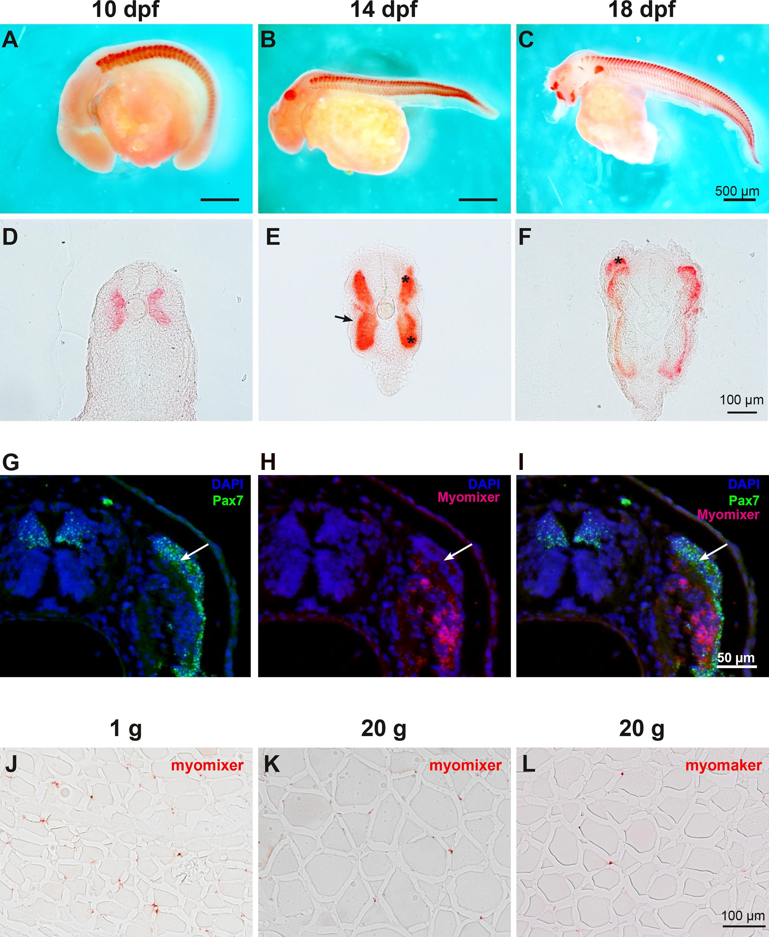
Fig. 3 Patterns of myomixer expression during embryonic development. (A-F) Embryos were analyzed by whole mounted in situ hybridization at day 10, 14 and 18 post fertilization. The corresponding vibratome section (35 µm) was presented for each stage. Asterisks indicate the dorsal and ventral domains of the myotome and arrowhead indicates the dermomyotome-like epithelium. (G-I) Double in situ hybridization for pax7 and myomixer of 17 dpf embryo sections. The nuclei are counter-stained with DAPI and arrowhead indicates the dermomyotome-like epithelium. (J-L) The expression of myomixer and myomaker in muscle of 1 g and 20 g trout was also studied using in situ hybridization on cross sections (7 µm).
Reprinted from Gene, 790, Perello-Amoros, M., Rallière, C., Gutiérrez, J., Gabillard, J.C., Myomixer is expressed during embryonic and post-larval hyperplasia, muscle regeneration and differentiation of myoblats in rainbow trout (Oncorhynchus mykiss), 145688, Copyright (2021) with permission from Elsevier. Full text @ Gene