
|
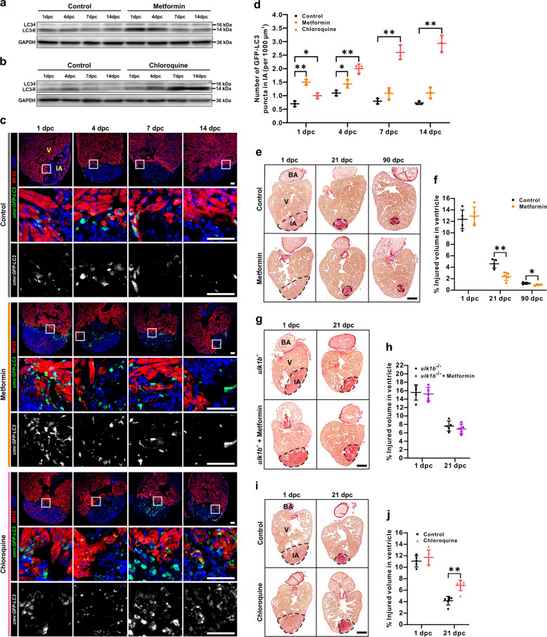
Fig. 2
a, b Western blot analysis was performed in cryoinjured hearts treated with or without 50 µM metformin (a) or 100 µM CQ (b) to detect the expression of LC3-I and LC3-II from 1 to 14 dpc. The protein from three hearts was loaded into each lane, and GAPDH was used as the loading control. c, d After cryoinjury, the heart of Tg(cmv:GFP-LC3) fish treated with or without 50 µM metformin or 100 µM CQ, were isolated, fixed, sectioned and dual-immunostained with anti-GFP (green) and anti-MF20 (red), after which they were stained with DAPI (blue). IA: injured area; V: ventricle. Scale bars: 50 µm (c). The number of GFP-LC3 puncta in the injured area at 1 dpc, 4 dpc, 7 dpc, and 14 dpc was quantified. The data are presented as mean ± SD, n = 3 hearts, *P < 0.05, **P < 0.01 vs control (d). e–j Following cryoinjury, the AB fish were either untreated (control) or else they were treated with 50 µM metformin for up to 90 days (e, f), or they were treated with 100 µM CQ for up to 21 days (i, j); the ulk1b mutant fish generated by TALEN were also either untreated (control) or else they were treated with 50 µM metformin for up to 21 days (g, h). Paraffin sections were then prepared and stained with Picro Sirius red. BA: bulbous arteriosus. Scale bars: 200 µm. The injured volume percentage was then quantified in the ventricle of the control and metformin (f, h) or CQ (j) treatment groups at the different time points. The data are presented as mean ± SD, n = 4 to 7 hearts, *P < 0.05, **P < 0.01 vs control.