
|
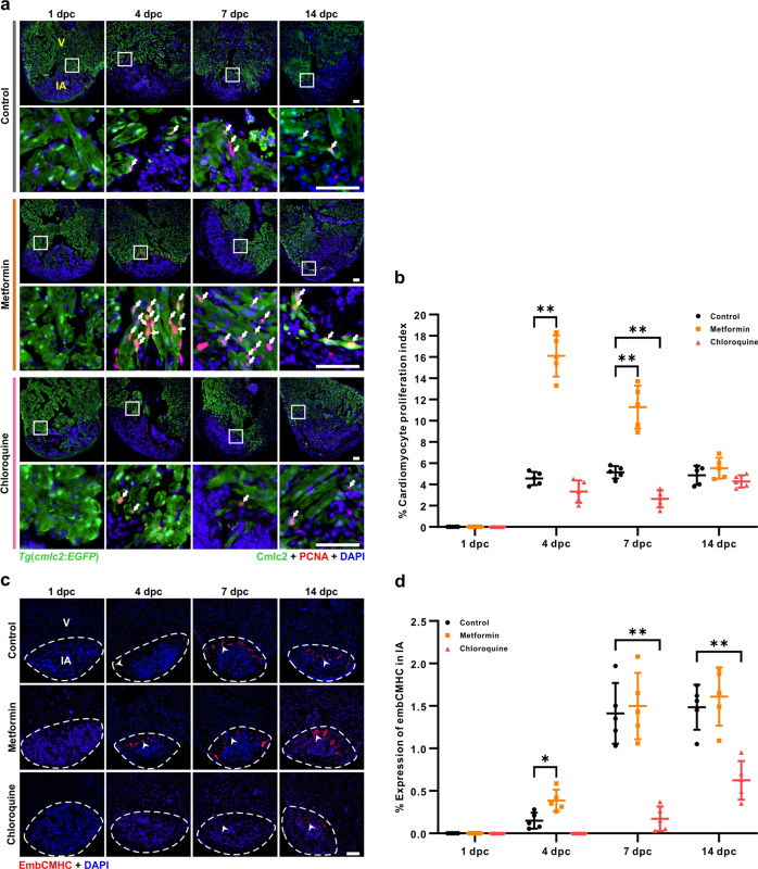
Fig. 3
a, b After cryoinjury, the Tg(cmlc2:EGFP) fish were maintained in untreated fish water (control) or in fish water containing 50 µM metformin or 100 µM CQ for the indicated days. The hearts were then isolated, fixed, sectioned and dual-immunostained with an anti-GFP antibody to identify the cardiomyocytes (in green) and an anti-PCNA antibody to identify proliferating cells (in red), after which they were co-stained with DAPI to label the nuclei (in blue). V: ventricle; IA: injured area. The white arrows show the proliferating cardiomyocytes. Scale bars: 50 µm (a). The proliferating cardiomyocytes in the untreated control, metformin and CQ treated fish at 1 dpc, 4 dpc, 7 dpc, and 14 dpc were quantified. The data are presented as mean ± SD, n = 5 hearts, **P < 0.01 vs control (b). c, d After heart cryoinjury, AB fish were maintained in untreated fish water (control) or in fish water containing 50 µM metformin or 100 µM CQ. The hearts were prepared, and immunostained with an anti-embCMHC (N2.261) antibody for the identification of embryonic cardiac myosin heavy chain (in red) and then they were labeled with DAPI. The arrowheads show the signal of embCMHC. Scale bar: 100 µm (c). The percentage of embCMHC positive cells in the control, metformin and CQ treated fish at 1 dpc, 4 dpc, 7 dpc, and 14 dpc was quantified. The data are presented as mean ± SD, n = 5 hearts, *P < 0.05, **P < 0.01 vs control (d).