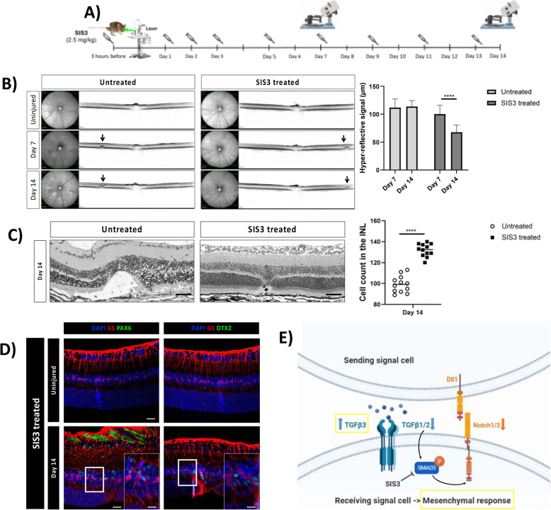
Pharmacological inhibition of p-Smad3 (SIS3) diminishes retinal damage. (A) SIS3 or PBS injections performed at 3 h before injury, daily during the first three days after injury, and then every other day until day 14. (B) IR (left) and OCT (right) images of the injury from a single animal at baseline, day 7 and 14. Arrows point to the central lesion depict the injury sites detected as hyper-reflective signal. Significant differences (****p < 0.001) between different time points were determined by two-tailed t-test (n = 12). (C) H&E-stained images of untreated and SIS3-treated retinas at day 14 after injury. Analyzed length was 100 μm, corresponding to the induced-laser burn. Significant differences in structural changes (****p < 0.0001) between untreated and SIS3 treated groups were determined by two-tailed t-test (n = 12). (D) Detection of PAX6 and OTX2 in GS+MCs in untreated and SIS3-treated mice. Shown are representative sections for GS (red) and PAX6 or OTX2 (green). (E) Schematic summary of molecular outcomes of SIS3 treatment in murine MCs. INL, inner nuclear layer; ONL, outer nuclear layer. Scale bar of the images equals 50 μm, while in the inserts corresponding to 150 μm
Acknowledgments
This image is the copyrighted work of the attributed author or publisher, and
ZFIN has permission only to display this image to its users.
Additional permissions should be obtained from the applicable author or publisher of the image.
Full text @ Mol. Neurodegener.
Your Input Welcome
Thank you for submitting comments. Your input has been emailed to ZFIN curators who may contact you if
additional information is required.
Oops. Something went wrong. Please try again later.